


Introduction
The brain is possibly the most complex of all biological systems. It allows us to receive, process and convert our surroundings into meaningful information. It also controls and regulates vital functions of the human body, including breathing, sleep pattern and appetite. This revision guide will explore the anatomy of this fascinating organ, including its vascular supply, embryological origin and much more.
Overview
The brain weighs approximately 1.5kg, accounting for 2% of our body weight. It comprises three parts; the cerebrum, cerebellum, and brainstem. The brain and spinal cord make up the central nervous system.
Cerebrum
The cerebrum is the largest part of the brain located within the bony cranium. It is divided into the left and right cerebral hemispheres, which are separated by the falx cerebri, a crescent-shaped fold of the dura mater.
Internally, the cerebrum consists of grey matter and white matter:
• Grey matter – Extends from the brain into the spinal cord and forms the surface of each cerebral hemisphere. The grey matter surrounding the cerebrum is known as the cortex of the brain.
• White matter – Found within the deeper tissues of the brain. It consists of glial cells and myelinated axons that transmit signals to the grey matter.
Externally, the cerebrum consists of sulci (grooves) and gyri (ridges) to increase its surface area, allowing for the presence of more neurons.
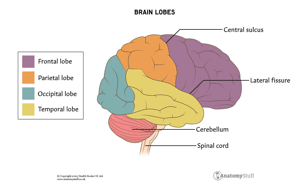
The cerebral hemispheres can be divided into lobes; frontal, temporal, parietal, and occipital.
| Lobes | Location | Function |
| Frontal | Anterior cranial fossa | · Higher cognitive functions, such as decision making
· Voluntary movement (motor cortex) · Motor component of speech (Broca’s area) |
| Temporal | Middle cranial fossa | · Hearing
· Sensory aspects of speech and memory |
| Parietal | Between the frontal and occipital lobe | · Sensation and perception
· Interpretation of sensory information |
| Occipital | Posterior cranial fossa | · Visual processing |
Cerebellum
The cerebellum is located within the posterior cranial fossa and plays a vital role in the regulation of movement and balance. It consists of 2 hemispheres and contains both grey and white matter, just like the cerebrum.
The cerebellum can be divided anatomically (anterior, posterior and flocculonodular lobes) and functionally (cerebrocerebellum, spinocerebellum and vestibulocerebellum).
Brainstem
The brainstem is located at the base of the brain and connects the cerebrum and cerebellum to the spinal cord. It comprises three different sections; the midbrain, pons, and medulla oblongata.
• Midbrain – The most superior of the three regions. The trochlear and oculomotor cranial nerves arise from here. The midbrain also includes many other important nuclei, including the substantia nigra, red nucleus and the dorsal raphe nucleus.
• Pons – The middle region consists of two main components; the ventral (basilar/basal) pons and the pontine tegmentum.
• Medulla Oblongata –The inferior portion which connects the brainstem to the spinal cord at the level of the foramen magnum, a large oval-shaped opening.
Deep structures
Above the brainstem lie many important structures:
• Thalamus – A paired structure that serves as the brain’s main relay station. The motor, limbic and sensory pathways (apart from olfaction) all pass through it.
• Hypothalamus – Located above the pituitary gland and below the third ventricle. It is made up of a collection of nuclei divided into three zones; lateral, medial and periventricular. The periventricular zone nuclei regulate the endocrine system.
• Pineal gland – A small endocrine gland located between the two cerebral hemispheres. It works closely with the hypothalamus to secrete melatonin which regulates the circadian rhythm (24-hour cycles that are part of the body’s internal clock).
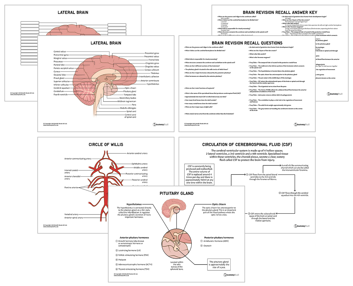
• Pituitary gland – Located within the sella turcica of the sphenoid bone. It consists of 3 main lobes – the anterior lobe and the posterior lobe. The hypothalamus regulates the pituitary gland’s secretion of many important hormones:
Anterior pituitary |
|
| Hormone | Function |
| Growth hormone (also known as somatotropic hormone or somatotropin) | Acts almost on every type of cell to stimulate skeletal growth. |
| Prolactin | Stimulates milk production and mammary gland (breast) growth. |
| Follicle-stimulating hormone (FSH) | Females – stimulates the growth and development of follicles.
Males – stimulates spermatogenesis. |
| Luteinising hormone (LH) | Females – triggers ovulation and stimulates the secretion of progesterone by the corpus luteum.
Males – stimulates testosterone secretion. |
| Thyroid-stimulating hormone (TSH) | Stimulates synthesis and secretion of thyroid hormones. |
| Adrenocorticotrophic hormone (ACTH) | Stimulates the secretion of adrenal hormones. |
Posterior pituitary |
|
| Hormone | Function |
| Oxytocin | Stimulates milk ejection from the breast during breastfeeding and uterine contractions during labour. |
| Antidiuretic Hormone (ADH) | Acts on the distal tubule and collecting ducts of the kidney to increase water reabsorption. |
Interesting fact!
The optic chiasm lies anterosuperior to the pituitary gland. This is an important part of the visual pathway where the optic nerves cross. Pituitary gland tumours may compress the chiasm and result in visual problems.
Meninges
The meninges refer to the membranous layers that cover and protect the central nervous system. From superficial to deep, the three layers are the dura, arachnoid and pia mater.
• The dura mater is a thick and dense fibrous membrane. It is composed of 2 layers: the periosteal layer and the meningeal.
Interesting fact!
In Latin, dura means hard or thick.
• The arachnoid mater is an avascular membrane involved with cerebrospinal fluid (CSF) metabolism.
• The pia mater is the deepest layer of the meninges. It is a thin and transparent layer that follows the contours of the sulci and gyri.
Ventricles and cerebrospinal fluid
The cerebral ventricular system is made up of 4 hollow spaces; 2 lateral ventricles, a 3rd ventricle and a 4th ventricle. Specialised tissue within these ventricles, the choroid plexus, secrete a clear, watery fluid called cerebrospinal fluid (CSF) to protect the brain from injury.
Cerebrospinal fluid is constantly being produced and reabsorbed. The entire volume of CSF is replaced around 4 times per day, and there is approximately 150ml at any one time within the brain.
Flow of CSF:
- CSF flows from the paired lateral ventricles to the 3rd ventricle through the foramen of Monro.
- CSF flows through the cerebral aqueduct into the 4th ventricle.
- CSF enters the subarachnoid space of the brain or spinal cord through the lateral and the median apertures.
- CSF is reabsorbed into the dural venous sinuses through small protrusions called arachnoid granulations.
Cranium
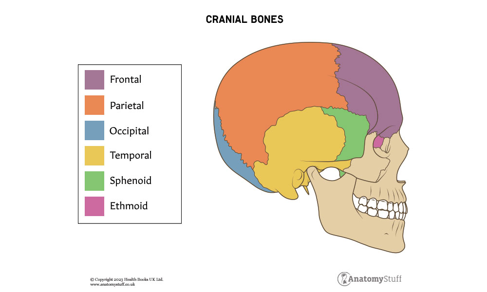
The skull consists of 22 bones, mainly cranial bones (8) and facial bones (14). The cranial bones act to protect the brain, along with the meninges. These bones include the frontal, parietal (2), temporal (2), sphenoid, occipital and ethmoid.
Cells of the brain
The central nervous system is made up of 2 basic types of cells; neurones and neuroglial cells (usually referred to as glial cells or glia).
Neurons are highly specialised cells which send and receive neurotransmitters. There are approximately 100 billion found within the brain and can be divided into three classes: sensory, motor and interneurons.
Glial cells support neurones by helping to maintain an appropriate chemical environment for neuronal signalling. There are three major types of glial cells: astrocytes, oligodendrocytes, and microglial cells.
| Type | Function |
| Astrocytes | Star-shaped cells which act as a “glue” to hold neurons together in their correct positions. |
| Oligodendrocytes | Form myelin sheaths around the axons of the CNS to provide insulation. |
| Microglial cells | Remove cellular debris by phagocytosis. They are very similar to macrophages. |
Unlike neurones, glial cells lack axons and dendrites. They are also smaller and more numerous throughout the brain.
Cranial nerves
The cranial nerves are a set of 12 paired nerves that arise directly from the brain. The first two nerves (olfactory and optic) arise from the cerebrum, whereas the remaining ten emerge from specific regions of the brainstem:
• Midbrain – Trochlear nerve (IV)
• Midbrain-pontine junction – Oculomotor (III)
• Pons – Trigeminal (V)
• Pontine-medulla junction – Abducens (VI), facial (VII), vestibulocochlear (VIII)
• Medulla oblongata – Glossopharyngeal (IX), vagus (X), accessory (XI), hypoglossal (XII)
Each cranial nerve can be described as being sensory, motor or mixed.
Blood supply
The brain receives blood from 2 sources: the internal carotid arteries (supply the anterior circulation) and the vertebral arteries (supply the posterior circulation). These systems connect at the Circle of Willis, a ring of vessels which allows for collateral blood flow between the anterior and posterior circulations of the brain. This highly significant safety mechanism helps to protect against ischaemia.
The brain’s venous circulation is different from other parts of the body as it does not correlate with the arterial supply. The deoxygenated blood empties into dural venous sinuses, collecting pools found between the layers of dura mater. The blood ultimately drains into the internal jugular vein to return to the right side of the heart.
Revision tip!
The Circle of Willis is tested a lot in exams, so familiarising yourself with the key arteries is important!
Embryology
Human brain development begins in the 3rd gestational week and continues postnatally. By the age of 6, the brain has reached approximately 90% of adult volume.
The cerebrum and brainstem arise from the rostral neural tube, which expands to form 3 primary brain vesicles:
• Prosencephalon – becomes the forebrain which later develops into the cerebral hemispheres
• Mesencephalon – becomes the midbrain
• Hindbrain/Rhombencephalon – becomes the hindbrain
As the brain develops, the 3 primary vesicles eventually give rise to 5 secondary brain vesicles: Telencephalon, Diencephalon, Mesencephalon, Metencephalon, and Myelencephalon, which each develop into a specific component of the nervous system.
Lymphatic drainage
For many years it was believed that the brain lacked a lymphatic system. However, recent studies have proven that a genuine and functional lymphatic vascular system does exist within the meninges. It is thought to play a vital role in the circulation and exchange of soluble contents between the CSF and the interstitial fluid.
You can discover more about the general anatomy of the body’s lymphatic system in our Lymphatic Revision Guide here.

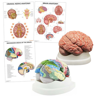
Related Products
View All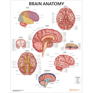

AnatomyStuff
Brain Anatomy Revision Worksheets (Interactive and Printable PDFs)
Now
£5.00
Inc VAT


