


Written by: Liz Paton, MSc
Elbow Anatomy Overview
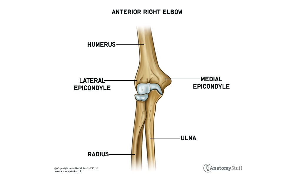
The elbow is the visible hinge joint where the upper arm articulates with the lower arm, it is made up of the humerus, ulna and radius bones.
Elbow Motion
Elbow range of motion includes flexion, extension, supination, pronation and neutral position.
Elbow flexion is where you move your forearm toward your body, such as lifting a dumbbell. Elbow extension is the opposite motion, where you move the forearm away from the body.
Supination is where you move your arm to face your palm upwards. Pronation is the opposite motion, where you move your arm to face your palm downwards. The neutral (or resting) position of the elbow is midway between pronation and supination with your thumb positioned upwards.
Bones and Joints of the Elbow
The elbow joint is a synovial hinge joint that joins the upper arm and lower arm, forming where the distal (lower end) humerus meets the proximal (upper end) radius and the ulna bones. These bones are all covered with cartilage (a tough, rubbery tissue that absorbs shock and allows joints to glide against one another).
The humerus bone is the longest bone in your arm, extending between the shoulder and elbow. The radius and ulna are the bones of the forearm, extending between the elbow and wrist.
The humeroradial joint is the articulation between the capitulum of the humerus and the fovea of the radius. The humeroulnar joint is the articulation between the trochlea of the humerus and trochlear notch of the ulna.
The proximal and distal radioulnar joints are the two locations of ulnar and radius articulation. They are involved with pronation and supination movements.
Muscles of the Elbow
There are several muscles that cross over the elbow joint that assist in movement and strength.
The muscles involved with flexion include the brachialis, brachioradialis and biceps brachii muscles. The biceps brachii consists of a short and long head. The triceps brachii muscle, consisting of a medial, lateral and long head, is responsible for extension. Often considered to be a part of the triceps brachii muscle on the posterior aspect of the elbow lies a small triangular muscle called the anconeus muscle which assists in extension.
The muscles involved with supination and pronation include the pronator teres, pronator quadratus, biceps brachii and brachioradialis muscles.
Ligaments of the Elbow
The ligament is a fibrous, tough, flexible connective tissue that connects and stabilise bones.
Two ligaments provide a substantial source of valgus (outward angulation) and varus (medial deviation) stability for the elbow: the medial and lateral collateral ligaments. The medial collateral ligament, located on the medial (inner) side of the elbow connects the ulna and humerus. The lateral collateral ligament (sometimes referred to as the radial collateral ligament) is positioned on the lateral (outer) side of the elbow and connects the radius and humerus.
The annular ligament surrounds the head of the radius and connects to the ulna at the radial notch. The quadrate ligament secures the proximal radius against the radial notch, contributing to elbow stability.
Free PDF Downloads
View All

Blood Supply of the Elbow
Arteries are blood vessels that carry oxygen-rich blood from your heart to tissues of your body.
The brachial artery is the major blood vessel of the arm that branches into the superior and inferior ulnar collateral arteries. The inferior ulnar collateral artery supplies the brachialis, biceps brachii and coracobrachialis muscles. The superior ulnar collateral artery provides the periarticular arterial anastomoses of the elbow joint. An anastomosis is a complex network of blood vessels to provide alternative routes if the vessels become damaged.
Nerves of the Elbow
There are three major nerves that run past the elbow called the median nerve, the ulnar nerve and the radial nerve.
The median nerve descends through the cubital fossa and gives off an articular nerve branch to the elbow joint. The cubital fossa is a small, innervated triangular area located on the anterior surface of the elbow of that forms the transition between the arm and forearm.
The ulnar nerve travels from the neck and runs behind the medial epicondyle of the elbow on the way to the hand. The radial nerve crosses the lateral epicondyle of the elbow on route from the neck to the hand.


