


Equine Anatomy
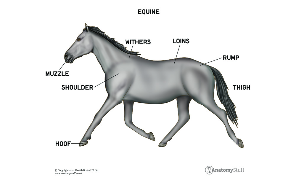
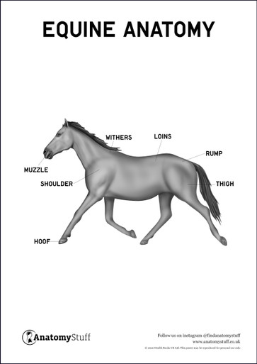
The Horse
The horse is a large mammal part of the Equidae family that has been domesticated for around 4,000 years. Most feral horses are descendants of horses that have been previously domesticated and therefore are not true wild horses. There is currently only true wild horse species remaining. Since horses have been domesticated for so long, they have been bred for specific purposes such as sports, leisure, labour and previously warfare.
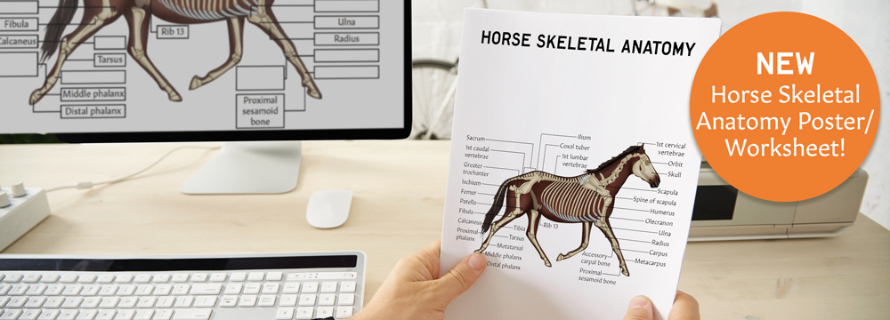
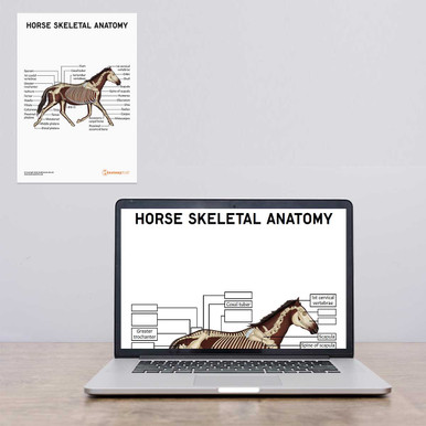
Horse Skeleton
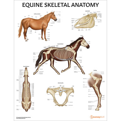
The skeletal system is split into two categories, the axial skeleton and the appendicular skeleton.
The axial skeleton consists of the skull, the vertebral column, the ribs and the sternum. The skull is made up of 37 fused bones which help to protect internal anatomy. The vertebral column is made up of 54 bones and extends from the base of the skull to the tail. There are 7 cervical vertebrae, 18 thoracic vertebrae, 6 lumbar vertebrae, 5 sacral vertebrae and between 15-20 caudal vertebrae. The horse has 18 ribs and protects vital internal organs.
The appendicular skeleton consists of the bones which form the forelimb, the hindlimb, the hip joint and the shoulder joint.
The forelimb consists of the scapula, the humerus, the radius, the ulna, the carpus, the metacarpals and the phalanges. The scapula provides an attachment point for various muscles with a large wing of cartilage attached distally. The humerus lies at a certain angle which allows for excellent shock absorption. The radius and ulna are fused together. The carpus consists of several bones which help with weight-bearing. The metacarpals are articulate with the lower row of the carpal bones and the phalanges articulate with the metacarpal bones. The proximal phalanx is known as the long pastern, the intermediate phalanx is known as the short pastern and the distal phalanx is known as the pedal bone.
The hindlimb consists of the pelvic girdle, the femur, the patella, the tibia, the fibula, the tarsus, the metatarsals and the phalanges. The pelvic girdle is made up of three bones, the pubis, the ischium and the ilium. The wings of the ilium are known as the sacral tubers. The femur attaches the pelvic girdle to the lower hindlimb. The patella, the lower femur and the proximal tibia and fibula make up the stifle joint. The tarsus, also known as the hock joint contains small bones which are positioned in three rows. The metatarsals and phalanges are the same as the forelimb.
Horse Muscles
The muscular system of the horse is made up of skeletal muscle, smooth muscle and cardiac muscle. These are some of the main skeletal muscles in the horse and their function.
Brachiocephalicus, sternocephalicus and splenius help with the movement of the head and neck. Brachiocephalicus also extends the shoulder joint. Movements of the shoulder joint are facilitated by the rhomboideus, trapezius, deltoids and latissimus dorsi.
Supraspinatus extends the forelimb and infraspinatus abducts and rotates the forelimb. Biceps brachii flexes the elbow and triceps brachii extends the elbow.
The superficial gluteal muscles provide extension and flexion of the hip. Biceps femoris, semitendinosus and semimembranosus help to extend the hip and flex the stifle joint. Gastrocnemius is important for flexing the stifle joint and extending the hock joint.
Horse Hoof
Horses are known as ungulates because they have hooves. The horse foot is a complex structure made up of soft tissue and a hard protective outer layer.
External Structures
The hoof wall is the outer layer that protects the internal structures and contains the protein keratin. This structure helps to support weight and absorb shock. It does not contain any nerves or vessels and requires regular trimming. The coronary band is situated just above the hoof and has a large blood supply which helps the hoof wall grow. The periople provides a protective outer layer to maintain moisture.
Underneath the Hoof
The sole is concave in shape and is similar in structure to the hoof wall. The frog is a tough V-shaped structure that helps with the circulation of the hoof and helps with shock absorption. The frog contains sensitive nerves which allow the horse to feel the surface they are walking on. The bulbs of the heel are located at the back of the foot and assists with promoting circulation. The bars provide strength to the heel and help to support the weight.
Internal structures
The internal structure of the hoof consists of bones including the navicular and pedal bone. The pedal bone provides shape to the hoof and absorbs weight. The navicular is located just behind the pedal bone and provides stability when walking on uneven ground. The digital cushion is a cartilaginous structure that provides shock absorption during weight-bearing. The lateral cartilages are attached to the pedal bone and assist with circulation. The corium is a vascularised layer of skin that supplies nutrients and blood to various parts of the hoof.
Free Download PDFs
View All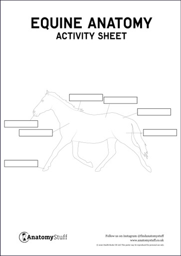
Related Products
View All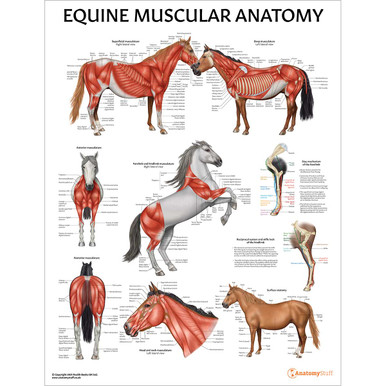
save
30%
AnatomyStuff
Horse Skeletal Anatomy (Interactive & Printable PDF)
Now
£3.50
Inc VAT
Was
£5.00
Inc VAT


