Written by: Liz Paton, MSc
Eye Anatomy Overview
The eye is the complex, spherical organ that allows us to see the world around us by reacting to light and working with our brain. The eye is a bilateral organ, meaning there are two eyes on each side of our face.
Anatomy of the Eye
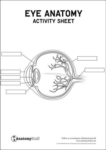
The eye is made up of three major layers; the fibrous tunic, vascular tunic and the retina.
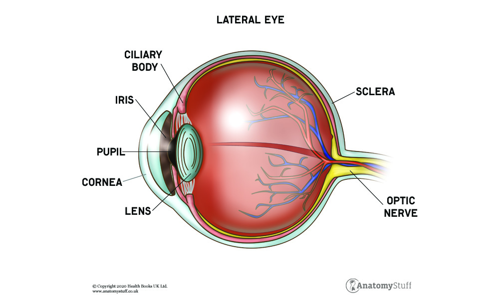
The fibrous tunic is the outermost layer of the eye composed of the sclera and cornea. The sclera is consistent with the cornea and is known as the “white of your eye”, maintaining the shape of the eye and serving as an attachment site to the extraocular muscles.
The vascular tunic (also known as the middle layer) is composed of the choroid, iris and ciliary body. The choroid serves as the major blood supply for the outer retina. The iris is the coloured part of your eye located around the pupil and between the cornea and the lens. The iris gets its colour from melanin. The ciliary body is an extension of the iris and produces the aqueous humour, the fluid that fills out eyeballs.
The retina (also known as the inner layer) is the layer of tissue that lines the back of the eye near the optic nerve. The retina is the part of the eye that enables vision by receiving light from the lens and transmitting signals to your brain for visual recognition.
Muscles of the Eye
Each eye consists of several important muscles responsible for movement, including the levator palpebrae superioris as well as six extraocular muscles and three intraocular muscles. The levator palpebrae superioris elevates your upper eyelid and maintains your eyelid’s position.
The extraocular muscles include the medial rectus, lateral rectus, superior rectus, inferior rectus, superior oblique and inferior oblique. The medial rectus muscle is the largest of the extraocular muscles and pulls the eye in a medial direction. The lateral rectus muscle moves the eye in a lateral direction, the superior rectus muscle pulls the eye upward and the inferior rectus muscle moves the eye downwards. The superior oblique muscle also moves the eye downwards. The inferior oblique muscle elevates and abducts the eye.
The intraocular muscles are the sphincter pupillae, dilator pupillae and the ciliary muscle. The sphincter pupillae (also known as the iris sphincter muscle) is located in the iris, encircling the pupil and constricting the pupil when it is exposed to bright light. The dilator pupillae muscle does just the opposite, increasing the size of the pupil to let more light in when the environment is dark. The ciliary muscle controls focus, changing the thickness and curvature of the eye’s lens.
How Vision Works
The way our vision works is very similar to how cameras work. First, light enters our eyes through the cornea (the front portion of our eye). It then passes through our pupil (the black part of our eye) and then hits the lens. The light then travels down the vitreous humour and then finally reaches the retina. The optic nerve (also known as cranial nerve II) transfers the visual information through electrical impulses.