


Written by: Liz Paton, MSc
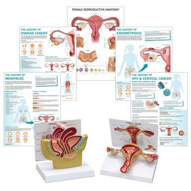
Female Reproductive System Overview
The Female Reproductive System produces the ova (eggs), sex hormones and helps to support the development of fertilised eggs. Important stages of reproduction and pregnancy occur within the structures of the female reproductive system.
External Organs of the Female Reproductive System
Vulva Anatomy
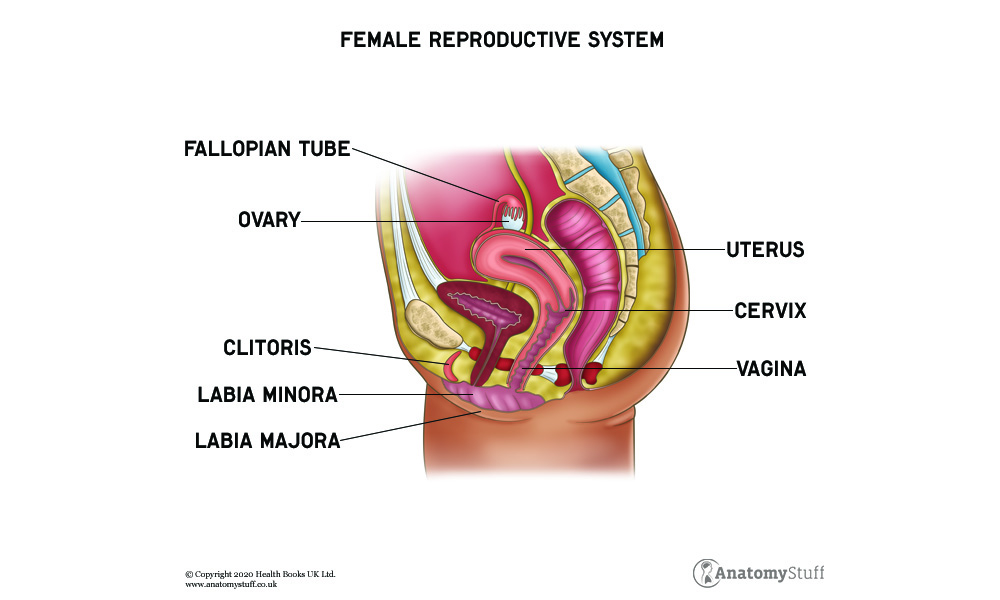
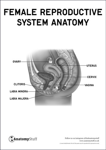
The vulva consists of the mons pubis, labia majora, labia minora, clitoris, vestibule, vestibular bulb and vestibular glands. The external female genitalia assist with reproduction, provide sexual pleasure and help to protect the internal female reproductive organs.
Internal Organs of the Female Reproductive System
Vagina Anatomy
The vagina is a muscular tube which expands and contracts. The walls of the vagina are lined with mucous membrane which protect the vagina and maintains lubrication. The vagina provides a passageway for various processes to occur such as giving birth, menstruation and sexual intercourse.
Uterus Anatomy
The uterus is the part of the reproductive system that receives the fertilised ovum and supports the development of the foetus during pregnancy. The uterus contains very strong muscles which expand and contract to help push the foetus out during childbirth.
There are three main parts of the uterus, the corpus, the isthmus, and the cervix. The corpus, also known as the body makes up the main part of the uterus. The base of the body is known as the fundus. The cervix is the lowest part of the uterus which connects the vagina to the uterus. The isthmus connects the corpus to the cervix.
Fallopian Tubes Anatomy
The fallopian tubes attach to either side of the uterus and connects the uterus to the ovaries. Fertilisation usually takes place in the fallopian tubes. The fertilised ovum known as a zygote is then transported to the uterus where implantation occurs. This is where the embryo attaches to the wall of the uterus.
Ovary Anatomy
The ovaries are located on either side of the uterus attached by various ligaments their function is to produce and stores ova. When an ovum is released into the fallopian tube it is known as ovulation. The ovaries also release hormones necessary for reproduction, menstruation and puberty.
Female Sex Hormones
Hormones help to send signals to parts of the body to maintain bodily functions. Oestrogen is a hormone mainly produced by the ovaries and helps with puberty, menstruation, pregnancy and menopause. Progesterone is another hormone produced by the ovaries and helps with thickening the lining of the uterus and helps with the early stages of pregnancy. Testosterone is produced by the adrenal glands and ovaries and helps to regulate the menstrual cycle. The luteinizing hormone and the follicle-stimulating hormone are present during puberty and during some of the menstrual cycle which helps to increase the production of oestrogen.
The Menstruation Cycle
The menstrual cycle is a natural cycle that occurs within the female reproductive system, where an ovum is released and the lining of the uterus sheds due to the rise and fall of oestrogen. An average cycle lasts around 28 days. There are four main phases of the cycle which include menstruation, the follicular phase, ovulation and the luteal phase.
Menstruation is when the thickened lining of the uterus sheds when fertilisation does not take place. Cells from the lining of the uterus and blood pass through the vagina. This period can vary in length but usually lasts around one week.
The follicular phase begins on the first day of menstruation and ends when ovulation occurs. The hypothalamus, which is part of the brain, causes the pituitary gland to release the follicle-stimulating hormone. As a result, the ovary produces follicles, each follicle contains an immature ovum. The production of follicles helps the lining of the uterus to thicken, which helps to prepare the uterus for pregnancy.
Ovulation occurs around day 14 of the cycle and is when a mature ovum is released from the ovaries. The follicle ruptures causing the ovum to be released and it is then transported into the fallopian tubes ready for fertilisation to occur.
The luteal phase is when the remains of the follicle turn into a structure called the corpus luteum and begins to release progesterone and oestrogen which helps maintain the thick lining of the uterus. If fertilisation occurs hormones are released to maintain the corpus luteum. When fertilisation does not occur the corpus luteum dies, causing the lining of the uterus to shed due to the fall in progesterone levels.
If you enjoyed our article on the female reproductive system and you’d like to learn about the male reproductive system then please click here.
Free Download PDFs
View All
Related Products
View All

AnatomyStuff
Female Reproductive System Anatomy & Pathology Collection
Now
£218.00
Inc VAT


