Written by: Liz Paton, MSc
Foot & Ankle Anatomy Overview
The foot and ankle joint consists of 28 bones and is a complex system which provides us with the ability to walk, run and jump. This part of the body is able to adapt to uneven terrain, provide shock absorption and allow for even and stable mobility during the gait (walking) cycle.
Motion of the Foot and Ankle
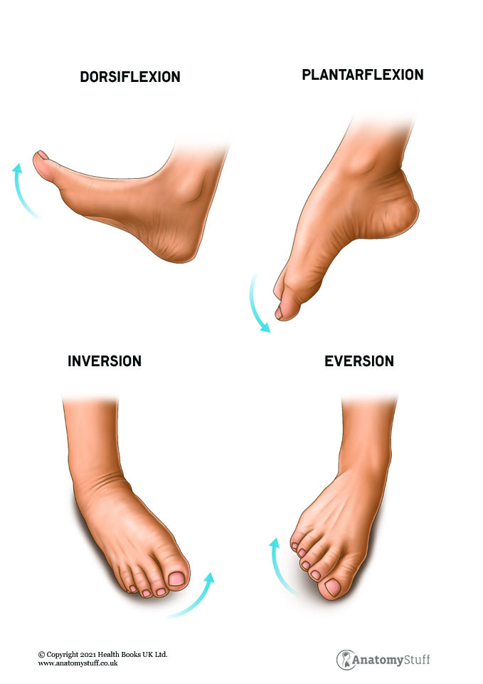
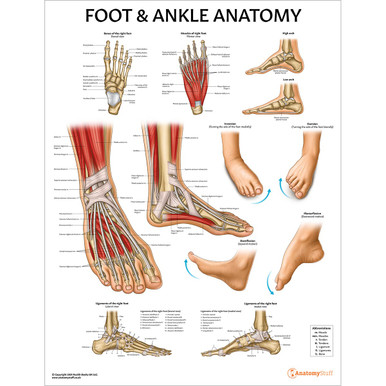
The ankle joint is also known as the talocrural joint. It allows the foot to dorsiflex (point upwards) and plantarflex (point downwards). This joint also allows the foot to adapt to uneven ground by providing side to side movement known as inversion (moving the foot inwards) and eversion (moving the foot outwards). The ankle also provides limited internal and external rotation.
Bones of the Foot and Ankle
Ankle
The ankle joint, called the talocrural joint, is formed when the end of the tibia and fibula (the two bones in the lower leg) meets a bone underneath called the talus. On the medial (inner) side of the tibia and the lateral (outer) side of the fibula are bony prominences called the malleolus. These bony landmarks are what you can feel when you touch your ankle.
Hind foot
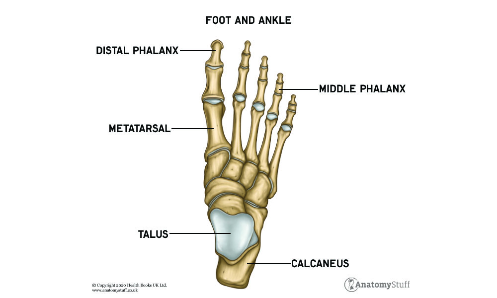
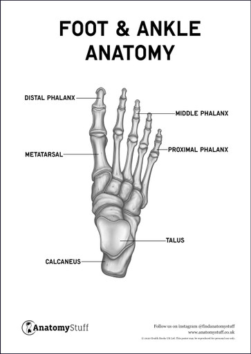
The hind foot connects the midfoot to the ankle and is made up of the talus and the calcaneus. The talus is an important bone because it has three articulations. As mentioned before, the talus articulates with the end of the tibia and fibula as well as the calcaneus and the navicular bone. This bone is important as it transfers body weight across the ankle joint.
The calcaneus is known as your heel bone and acts as a lever during dorsiflexion and plantarflexion of the foot. This bone has an important role in weight bearing and providing stability during walking. The calcaneus articulates with the talus and a bone called the cuboid.
Midfoot
The bones of the midfoot are called the navicular, cuboid, lateral cuneiform, medial cuneiform and intermediate cuneiform. The midfoot connects the forefoot to the hindfoot and helps to form the arches of the feet.
Forefoot
The forefoot consists of the metatarsal bones and the phalanges. The big toe is called the hallux and is unique because it contains two phalanges and one metatarsal bone. The hallux also has two small bones which sit at the base of the toe called sesamoid bones. These small bones help the toe push off the ground. The other toes are made from one metatarsal and three phalanges.
Joints of the Foot and Ankle
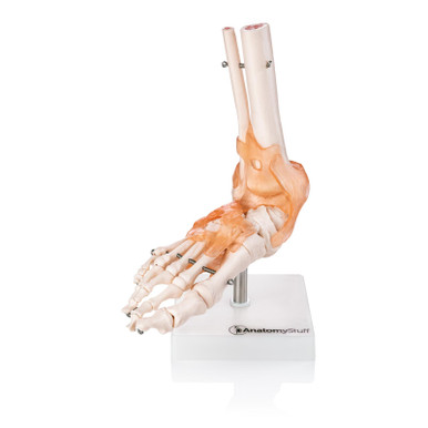
The bones of the foot articulate with each other through synovial joints. A synovial joint allows for smooth motion between adjacent bones. There are intertarsal joints, tarsometatarsal joints, metatarsophalangeal joints and interphalangeal joints.
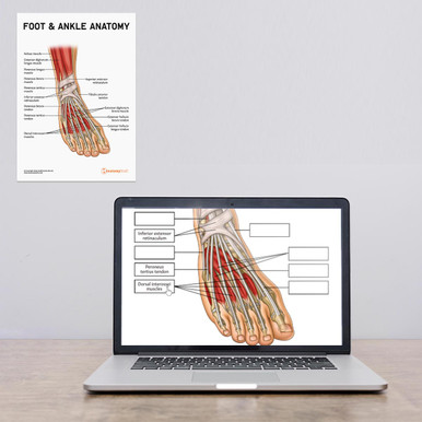
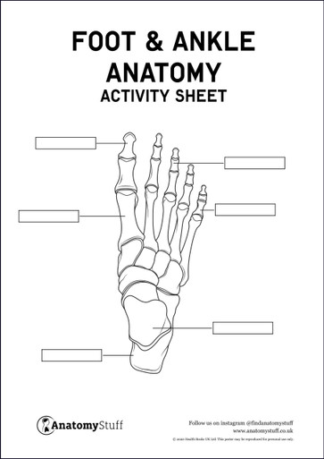
Pair this with our Foot and Ankle Anatomy Activity Sheet for some extra revision.
Muscles of the Foot and Ankle
There are three compartments of the lower leg which contain muscles that insert into the foot. These are all extrinsic muscles because these muscles are located in the lower leg.
Extrinsic muscles of the foot and ankle
Anterior
The anterior (front) compartment contains the tibialis anterior, extensor hallucis longus, extensor digitorum longus and peroneus tertius. The main job of this compartment is to dorsiflex the ankle. However, the extensor hallucis longus and extensor digitorum longus also extend the toes. Peroneus tertius everts and externally rotates the foot.
Lateral
The lateral (outer) compartment contains peroneus longus and peroneus brevis which evert and plantarflex the foot.
Posterior
The superficial posterior (rear) compartment contains the gastrocnemius, soleus and plantaris. The gastrocnemius inserts into the calcaneus where is becomes the Achilles tendon. This tendon assists the gastrocnemius and the soleus with plantarflexion and also has a role in stabilising the foot and ankle.
There is also a deep posterior compartment which contains the tibialis posterior, flexor digitorum longus and flexor hallucis longus. The primary role of muscles in the posterior compartment is to plantarflex the ankle.
Intrinsic Muscles of the Foot and Ankle
Intrinsic muscles of the foot and ankle are muscles which are located within the foot itself. These muscles can be split into dorsal (top of the foot) and plantar (base of the foot) muscles.
Dorsal
The dorsal muscles consist of extensor digitorum brevis which extend the toes, four dorsal interossei muscles, and extensor hallucis brevis which extends the big toe.
Plantar
The first layer of muscles includes the abductor hallucis, which abducts the big toe, the flexor digitorum brevis, which flexes the second to fifth toe and the abductor digiti minimi, which abducts the fifth toe.
The second layer includes quadratus plantae which flex the furthest phalanges and the four lumbricals which flex the joints between the phalanges and the metacarpals.
The third layer includes hallucis brevis which flexes the big toe, the oblique and transverse head of adductor hallucis which adducts the big toe and flexor digiti minimi brevis which flexes the fifth toe.
The fourth layer includes three plantar interossei muscles which adduct the toes.
Ligaments
Ligaments of the Ankle
A ligament is fibrous connective tissue which attaches bone to bone. The ligaments within the ankle joint stabilise the ankle and resists against excessive movement and rotational stress.
The medial collateral ligament attaches to the medial malleolus and the calcaneus, talus and navicular.
The lateral collateral ligament consists of three separate ligaments; the anterior talofibular ligament, the posterior talofibular ligament and the calcaneofibular ligament.
Ligaments of the Foot
The Lisfranc ligament stabilises the bones in the midfoot. The inter-metatarsal ligaments keep the metatarsals moving together.
Arches of the foot
The arches of the foot play an important role in helping with weight bearing, shock absorption and propulsion during everyday activities. They are made up of bones, ligaments and muscles which support provide support when walking. There arches consist of the medial longitudinal arch, the lateral longitudinal arch and the transverse arch.
Blood Vessels of the Foot and Ankle
Arteries are blood vessels that carry oxygen-rich blood from your heart to tissues of your body.
One branch of the popliteal artery gives rise to the anterior tibial artery, which supplies blood to the anterior compartment of the leg. When this artery reaches the ankle, it is called the dorsalis pedis artery.
A separate branch of the popliteal artery is the posterior tibial artery which supplies blood to the superficial and deep compartments of the posterior of the leg.
The fibular artery is a branch of the posterior tibial artery that supplies the lateral compartment of the leg.
Nerve Supple of the Foot and Ankle
The nerves in the foot allow you to feel the world you walk around by transmitting signals to your brain. The superficial peroneal nerve, deep fibula nerve, tibial nerve, sural nerve and saphenous nerve all supply innervation to the foot. These nerves originate from the sciatic nerve, a large nerve branch that branches from the lower back, through the hips and buttocks, and down the leg to the foot.
Free PDF Downloads
View AllRelated Products
View All