Written by: Liz Paton, MSc
Shoulder Anatomy Overview
The shoulder is a flexible joint made up of three bones that connects our upper limb to our torso and allows us to move our arm in several directions.
Shoulder Joints
The shoulder is made up of three total joints, making it the most flexible joint in our body.
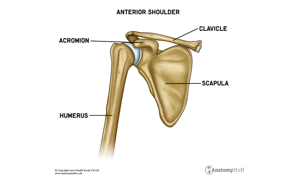
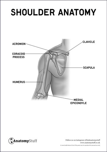
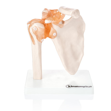
The glenohumeral joint is where the humerus (the upper arm bone) connects to the scapula (the shoulder blade) forming a ‘ball-and-socket’ for range of motion. Situated between the ‘ball-and-socket’ is cartilage that keeps the joint in place whilst maintaining flexibility, called the glenoid labrum.
The acromioclavicular joint forms the junction between the clavicle (the collarbone) and the scapula. This is a plane synovial joint, also known as a gliding joint (joints between flat articular surfaces).
The sternoclavicular joint is where the medial aspect of the clavicle and manubrium of the sternum meet just below the neck, anchoring the clavicle and allowing it to move.
Shoulder Motion
The shoulder joint allows us to move our arms in several different motions.
Shoulder flexion is when you move your arm away from its resting position and up toward your head, involving the deltoid, pectoralis major and coracobrachialis muscles. Extension is when you move your arm behind your body, involving the latissimus dorsi, teres major, teres minor and deltoid muscles.
Abduction is when you move your arm away from your body, as the name suggests, and form a “T” shape with your body. Adduction is where you move your arm back toward the centre of your body.
Circumduction is when you move your arm moves in a circular motion; using a combination of flexion, abduction, extension and abduction.
Medial rotation (also known as internal rotation) is where you move your arm inward toward your body. Lateral rotation is when you move your arm away from your midline.
Muscles of the Shoulder
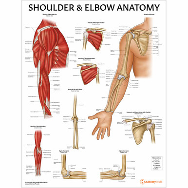
Holding the shoulder joints together is a series of muscles and ligaments. The muscles of the shoulder are divided into two groups depending on where they insert and originate from called the extrinsic and intrinsic muscle groups.
Extrinsic muscles
Extrinsic muscles originate from the torso and insert into the humerus, responsible for the movement of the upper limb. The extrinsic muscles include the trapezius, latissimus dorsi, levator scapulae, rhomboid major and rhomboid minor.
Intrinsic muscles
The intrinsic muscles originate from the scapula and/or clavicle and attach to the humerus.
The supraspinatus and infraspinatus muscles are the upper and lower muscles of the shoulder blade. Below these, you’ll find the teres minor and the subscapularis muscles. These four muscles make up the rotator cuff.
Sitting above these is the powerful deltoid muscle, a thick, triangular-shaped muscle which primarily helps you abduct (move away from you) your shoulder and prevents subluxation or dislocation of the humerus. The teres major is a medial rotator and an adductor of the humerus.
Ligaments of the Shoulder
The ligament is a fibrous, tough, flexible connective tissue that connects and stabilise bones. The ligaments in the shoulder get their names from the bones they attach. The glenohumeral ligaments are three ligaments that join the glenoid fossa to the humerus called the superior, middle and inferior glenohumeral ligaments. The acromioclavicular ligament attaches the acromion (a bone process of the scapula) to the clavicle. The coracoclavicular ligament attaches the coracoid process of the scapula to the clavicle. The coracohumeral ligament is a strong ligament connects the coracoid process of the scapula to the greater tubercle of the humerus.
Tendons of the Shoulder
The tendon is a fibrous connective tissue that connects muscle to bone. Some of the major tendons in the shoulder include tendons of the rotator cuff and the biceps tendons. The long head of the biceps tendon attaches to the glenoid and the short head of the biceps tendon attaches to the coracoid process.
Shoulder Blood Supply
Arteries are blood vessels that carry oxygen-rich blood from your heart to tissues of your body. The subclavian artery arises from the heart and branches off to the axillary artery which provides oxygenated blood to the muscles and bones of the shoulder. The brachial artery branches off from the axillary artery and supplies the shoulder with deoxygenated blood and then branches off to the forearm and hand.
Veins return deoxygenated blood towards your heart. The cephalic vein and subclavian vein run opposite each other, passing through the shoulder continuing to the axillary vein. The axillary vein drains from the lateral aspect of the armpit into the subclavian vein on the way to the heart.
Nerves of the Shoulder
Nerves are responsible for carrying messages from the hand to your brain for sensation, reflexes and movement.
All of the nerves that supply the shoulder joint arise from the brachial plexus, a major network of nerves that sends signals from the spinal cord (C5 – T1) to the shoulder and the rest of the upper limb.
The axillary nerve arises from the C5 and C6 nerve roots and innervates the teres minor and deltoid muscles. The suprascapular nerve also arises from the C5 and C6 nerve roots and innervates the supraspinatus and infraspinatus muscles. The lateral pectoral nerve arises from the C5-C7 nerve roots and innervates the pectoralis major muscle. The long thoracic nerve arises from C5-C7 nerve roots and innervates the serratus anterior muscle, which anchors the scapula to the chest wall. The musculocutaneous nerve arises from the C5-C7 nerve roots and innervates the biceps muscle.
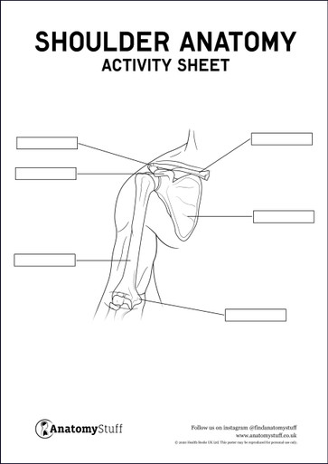
For more revision materials take a look at our Shoulder Anatomy Revision Worksheets.


