


Written by: Liz Paton, MSc
Skull Anatomy Overview
Made up of 22 bones, the skull plays a very important role in protecting the brain from injury while also supporting various structures of the face.
Bones
The bones of the skull can be split into two categories the neurocranium and the viscerocranium. There are eight neurocranium bones that protect the brain and fourteen viscerocranium bones that form the structure of the face.
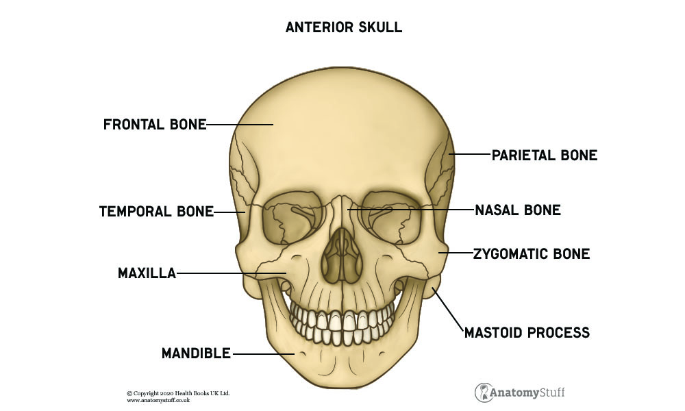
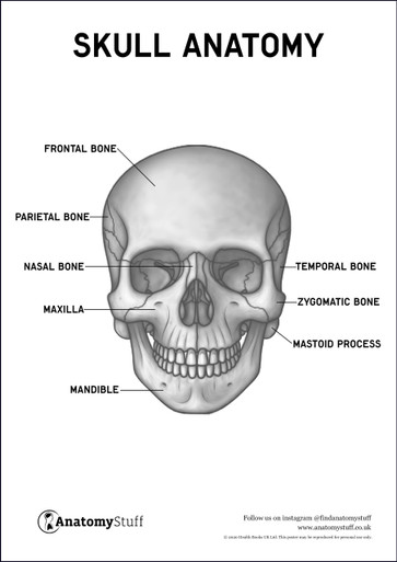
The neurocranium consists of the calvaria and the skull base. The calvaria is the skull cap and is the top part of the skull which protects the brain which is made from the frontal bone, the parietal bones, the temporal bones and the occipital bone. The frontal bone sits at the front of the skull where your forehead is located. There are two parietal bones that sit either side of the top of the skull and two temporal bones that sit just underneath each parietal bone. The occipital bone forms the back of the skull. The skull base is the lower part of the neurocranium and also protects the brain where the sphenoid and ethmoid bone are both located.
The viscerocranium provides attachment sites for all of the muscles we use to make facial expressions. The mandible joins the temporal bone on either side to form our jaw. The two maxilla bones are located at the front of our face which sits above our jaw. The vomer is located inside our nasal cavity along with the two nasal conchae. The two nasal bones form the top of the nasal cavity. The two zygomatic bones form our cheek bones. The two lacrimal bones sit near our eye socket known as the orbit and houses a lacrimal sac that forms our tear ducts. The two palatine bones help to form the oral cavity.
Sutures
A suture is a strong fibrous joint which holds the bones of the skull together. The joints of the skull are made from cartilage which is a tough but flexible tissue that covers bone surfaces. The coronal suture joins the frontal bone with the two parietal bones. The sagittal suture joins the two parietal bones where they meet in the centre. The lambdoid suture joins the parietal bones with the occipital bone. The squamosal suture joins the temporal bones with the parietal bones.
Muscles
The muscles of mastication help us with the start of digestion which involves chewing food. These muscles include the masseter, the temporalis, the medial pterygoid and the lateral pterygoid.
The muscles of the face provide a role in facial expression, vision and speech. The facial muscles can be split into five categories: the muscles of the mouth, the muscles of the nose, muscles of the eyelids, muscles of the cranium and neck, and the muscles of the ear.
Muscles of the mouth: orbicularis oris, buccinator, levator laii superioris, depressor labii inferioris, levator labii superioris alaeque nasi, mentalis, risorius, levator anguli oris, zygomaticus major and zygomaticus minor.
Muscles of the nose: nasalis and procerus
Muscles of the eyelid: orbicularis oculi, corrugator supercilii
Muscles of the cranium and neck: occipitofrontalis and platysma
Blood supply
The main blood supply is from the carotid arteries which supply the front of the skull and the vertebral arteries which supply the back of the skull.
The common carotid artery branches into the internal and external carotid arteries. The internal carotid artery helps to supply the structures within the cranium. The external carotid artery supplies blood to the skull bones and the meninges which is a membrane that covers the brain for protection.
The internal carotid artery and the vertebral arteries come together to form an anastomosis. This is a complex network of blood vessels that provide an alternative route for blood to move through if injury to the brain occurs. This ensures that vital structures are provided with blood even if part of the network becomes damaged. This particular anastomosis is called the circle of Willis.
The internal and external jugular veins are responsible for transporting deoxygenated blood back towards the heart.
Nerves
The skull contains bony structures called foramina (small holes). The foramina allow various nerves and blood vessels to pass through. The cranial nerves pass through foramina in the skull to innervate the rest of the body. The cranial nerves consist of the olfactory nerve, optic nerve, oculomotor nerve, trochlear nerve, trigeminal nerve, abducens nerve, facial nerve, vestibulocochlear nerve, glossopharyngeal nerve, vagus nerve, accessory nerve and the hypoglossal nerve.
The nerves of the skull, scalp and face receive innervation from the branches of the trigeminal nerve which include the ophthalmic, maxillary and mandibular nerves. The branches of the occipital nerve supplies innervation to the back of the skull and scalp. Most of the muscles of facial expression are innervated by the facial nerve.
Take a look at our Skull Anatomy Revision Worksheets for the best revision experience.
Free Download PDFs
View All
Related Products
View All


save
30%
AnatomyStuff
Skull Anatomy Poster / Worksheet (Interactive & Printable PDF)
Now
£3.50
Inc VAT
Was
£5.00
Inc VAT


