Introduction
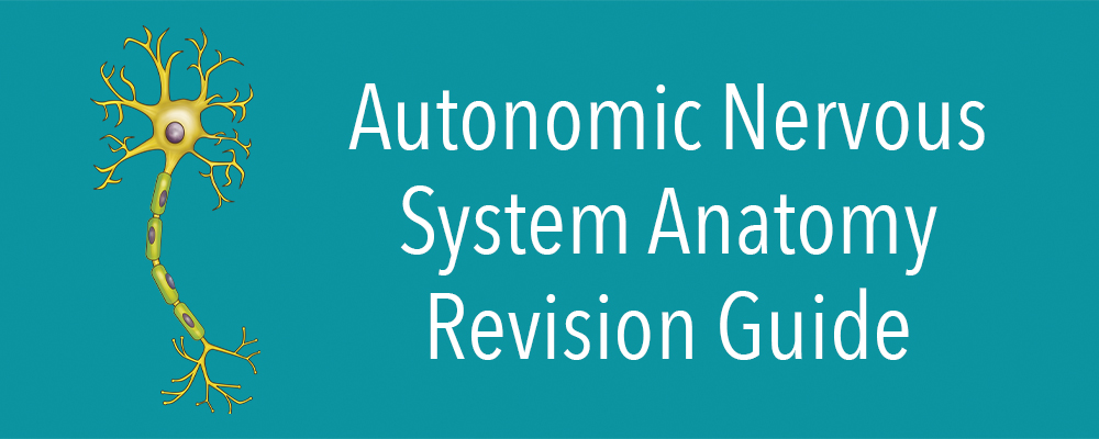
The nervous system is the body’s main controlling, regulatory, and communicating system. It consists of the central nervous system (CNS) and the peripheral nervous system (PNS). The autonomic nervous system (ANS) is a subcomponent of the peripheral nervous system (PNS) that regulates involuntary physiologic processes, including blood pressure, heart rate, respiration and digestion. It can be further divided into the sympathetic, parasympathetic, and enteric systems. This revision guide will explore the anatomy of the autonomic nervous system, including the functions of the sympathetic, parasympathetic and enteric nervous systems.
Recap!
The nervous system can be divided into CNS and PNS:
• CNS consists of the brain and spinal cord
• PNS consists of everything outside the brain and spinal cord
The PNS consist of the following:
• Autonomic nervous system (ANS): Controls involuntary bodily functions
• Somatic nervous system: Controls voluntary movements via skeletal muscles. It consists of both afferent (sensory) and efferent (motor) nerves
The autonomic nervous system can be divided into:
• Sympathetic nervous system (SNS)
• Parasympathetic nervous system (PNS)
• Enteric nervous system
Autonomic Nervous System (ANS)
The autonomic nervous system functions without conscious control to regulate physiologic functions necessary for human survival. As discussed above, it consists of three distinct divisions: sympathetic, parasympathetic and enteric. Most organs in the human body are controlled primarily by either the sympathetic or the parasympathetic division, which have opposite, antagonistic effects. For example, the sympathetic division increases blood pressure while the parasympathetic division decreases it. Therefore, in essence, the two divisions work together to ensure that the body responds appropriately to different situations.
Effects of the ANS include:
• Perfusion through regulation of the heart rate and blood pressure
• Homoeothermic role through sweating control and shivering
• Processing of nutrients through control and coordination of different parts of the gut
• Urinary motility
• Pupil movement
Interesting fact!
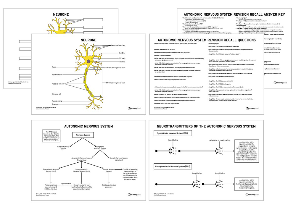
The ANS is also known as the visceral nervous system. The word ‘viscera’ refers to the internal organs.
Sympathetic Nervous System
The sympathetic nervous system promotes energy expenditure while inhibiting digestion. It is also known as ‘fight or flight’ because it originally evolved as a survival mechanism, enabling us to react quickly to life-threatening situations by either ‘fighting’ or ‘fleeing’ during times of imminent danger. The effects of the sympathetic nervous system include:
• Increase in heart rate and cardiac muscle contractility
• Decreased GI motility
• Decreased urine output
• Pupil dilation (ciliary muscle relaxes)
• Increased secretions from sweat glands
It originates within the thoracic and lumbar segments of the spinal cord, more specifically, T1-T12 and L1-L3. The sympathetic pathway can be divided into three following components:
• The preganglionic neurons
• The sympathetic ganglia
• The postganglionic neurons
The preganglionic neurons synapse onto postsynaptic neurons found within the sympathetic ganglia. The neurotransmitter (a chemical substance which is released at the end of a nerve fibre) used at this junction is acetylcholine which activates nicotinic receptors. The postganglionic neurons then release the neurotransmitter adrenaline or noradrenaline.
Interestingly, the only exception to the postganglionic release of adrenaline or noradrenaline is the sympathetic innervation of sweat glands and the arrectores pili muscles (small muscles attached to hair follicles). They use acetylcholine as their postganglionic neurotransmitter.
Parasympathetic Nervous System
The parasympathetic nervous system aims to conserve energy and regulate bodily functions like digestion and urination. Therefore, it is often known as the ‘rest and digest’ system rather than ‘fight and flight’. It is composed of pre and postganglionic neurons that act on effector organs, just like the sympathetic system.
The preganglionic neurons arise from both the brainstem nuclei and the sacral spinal cord (specifically S2-S4). They are much longer than the neurones found within the sympathetic nervous system, and the postganglionic neurons are very short. When the preganglionic neurons synapse with the postganglionic neurons near the effector organs, the short postganglionic axons then relay signals to the cells of the effector organs.
It is important to be aware that the parasympathetic neurons arising from the brainstem leave the central nervous system (CNS) through cranial nerves. Cranial nerves carrying parasympathetic functions include:
• Oculomotor nerve (III)
• Facial nerve (VII)
• Glossopharyngeal nerve (IX)
• Vagus nerve (X)
Interesting Fact!
You can remember which cranial nerves are involved in the parasympathetic nervous system by the year ‘1973’ – the 1 representing 10 (vagus), the 9 representing glossopharyngeal, the 7 representing facial and the 3 representing oculomotor.
The functions of the parasympathetic include:
• A decrease in heart rate and contractility of cardiac muscle
• Constriction of the ciliary muscle and the pupil for near vision
• Increased secretion by lacrimal glands and salivary glands
• Increased gut motility
• Contraction of the detrusor muscle with the relaxation of urethral sphincters
The PNS uses acetylcholine as its neurotransmitter for both pre and postganglionic neurons activating muscarinic receptors.
Enteric Nervous System
The enteric nervous system is responsible for regulating digestive processes within the gastrointestinal tract. It is made up of over one hundred million neurones found within the lower oesophagus all the way to the rectum. It also includes two plexuses (myenteric and submucosal) and their associated ganglia. Interestingly, the enteric nervous system can function completely independently from the rest of the nervous system.
Recap!
Ganglia are bundles of neuronal bodies found in the PNS. They interconnect with other ganglia to form a complex ganglia system known as a plexus.
The myenteric plexus is also known as the Auerbach plexus. It supplies motor innervation to the muscularis externa layer of the digestive tract and therefore controls peristalsis. It is partly controlled by the vagus nerve, and the main neurotransmitters involved are acetylcholine and nitric oxide.
The submucosal nerve plexus is also known as the Meissner plexus. Unlike the myenteric plexus, which is located in the muscularis externa, this plexus is found within the submucosa of the digestive tract. It innervates cells in the epithelial layer and the smooth muscle of the muscularis mucosae.
Development
The autonomic nervous system forms through the migration of neural crest cells. These are stem cells that originate from the neural tube and migrate to different embryonic tissues to give rise to a wide variety of cell types.
Autonomic dysfunction
A wide range of factors can cause autonomic dysfunction, which means problems with the autonomic nervous system. Some of these factors include:
• Autoimmune conditions – Guillain-Barre and rheumatoid arthritis
• Metabolic/ Nutritional – diabetes and vitamin B12 deficiency
• Infections – human immunodeficiency virus (HIV) and Lyme disease
• Toxins – alcohol and chemotherapy
• Medications –
• Dopamine antagonists
• Atypical antipsychotics
• Calcium channel blockers
• Selective serotonin receptor reuptake inhibitors
Related Products
View All