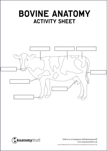
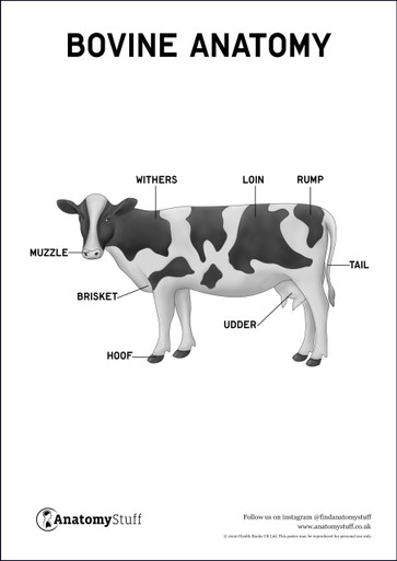
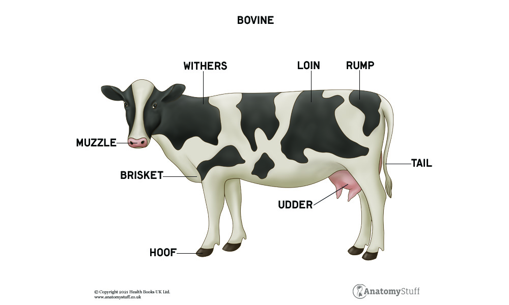
Cow Anatomy
Humans have used domesticated bovines for around 10,000 years to increase the supply of fresh food and materials rather than rely on hunting. Cows are domesticated farm animals and are one of the commonly known types of bovines. Cows are quadrupedal animals which means that they walk on four legs. They are also known as ungulates because they have hooves.
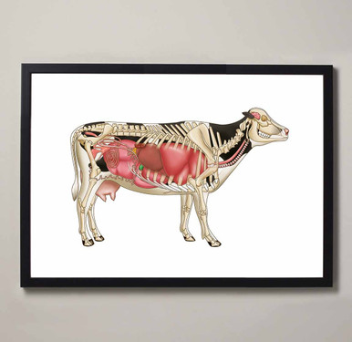
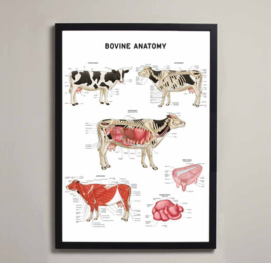
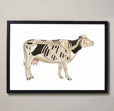
Cow Skeleton
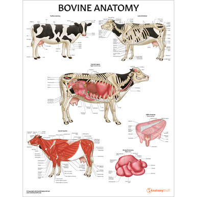
The cow skeleton can be divided into axial, appendicular and splanchnic. The axial skeleton consists of the skull, the ribs and the vertebral column. The skull is similar to a dog’s skull but is more elongated. The vertebral column starts at the base of the skull and runs horizontally along the body. There are seven cervical vertebrae, thirteen thoracic vertebrae, six lumbar vertebrae, five fused sacral vertebrae and 18-20 coccygeal vertebrae. The cow has thirteen pairs of ribs that attach to the spinal column and the sternum on either side.
The appendicular skeleton consists of two forelimbs and two hindlimbs. The forelimb is made up of a scapula, humerus, radius, ulnar, the capus, metacarpals and phalanges. The hindlimb consists of the pelvic girdle which includes the ilium, ischium and pubis. The tuber coxae sit on either side of the hip bone and determines the angulation of the femur in relation to the pelvis. The femur and the tibia form the stifle joint (knee joint) and the patella sits over this joint. The rest of the hind limb consists of the fibula and the tarsus which is made up of five small bones.
The splanchnic skeleton are the bones located in the heart called ossa cordis. These bones are not attached to any other bones and their function is to provide strength to the soft tissue.
Cow Muscles
The muscles of the cow are the same as the muscles of the horse. However, due to selective farming, domesticated cows are now bred to produce the best cuts of meat and therefore the size of the muscles are larger in comparison to the horse.
Muscles of the forelimb
The muscles which join the forelimb to the trunk include the trapezius, brachiocephalic muscle, omotransversarius, latissimus dorsi, pectoral muscles, and the rhomboids. These muscles help to transfer weight from the body to the forelimbs.
The muscles of the shoulder include the deltoid muscles, teres major, teres minor, supraspinatus, infraspinatus, subscapularis and coracobrachialis. These muscles provide flexion and stability to the shoulder joint.
The elbow joint extensors include the triceps brachii and the tensor fasciae antebrachii. The flexors of the elbow joint are the biceps brachii and brachialis. There are six extensor muscles and five flexor muscles of the carpal and digital joints of the lower limb.
Muscles of the hindlimb
The biceps femoris muscle has various points of insertion and its job is to extend and abduct the hindlimb. The semimembranosus and semitendinosus help to extend the hip and the stifle joint. The medial compartment of the thigh helps with flexion and extension of the hip and adduction of the hindlimb.
The stifle joint is controlled by the quadriceps muscle. There are four muscles within this compartment which include vastus lateralis, vastus medias, vastus intermedius and the straight muscle, they extend the stifle joint and flex the hip joint. The popliteal muscle flexes the stifle joint and pronates the lower leg.
Cow Digestive System
The cow is known as a ruminant animal which means they have a special digestive system that has adapted to process plant-based foods. The stomach has four separate compartments which each possess their own function.
Digestion begins at the mouth where saliva contains an enzyme that helps to start the initial breakdown of food. When the food has been swallowed it enters the oesophagus which contains muscles that help transport food up to the mouth or down to the stomach. The oesophagus has this function because cows regurgitate their food where it is broken down further by the mouth.
Once the food has been broken down properly it will reach the rumen which is the first compartment of the stomach. This compartment stores chewed vegetation which is known as cud. Once the rumen absorbs some nutrients from the cud it is transported over to the reticulum. This compartment holds dense material that cannot be digested such as rocks. This material is then regurgitated as it cannot be digested. The omasum is the next compartment that helps to absorb nutrients and water. The abomasum is the last compartment that contains acid to help break down the food further.
The small intestine consists of the duodenum, jejunum and the ileum which help to absorb nutrients and prevents the backflow of digested substances. The cecum sits between the small intestine and the large intestine which helps with the breakdown of food. The large intestine absorbs any leftover water.