


![]()
Introduction
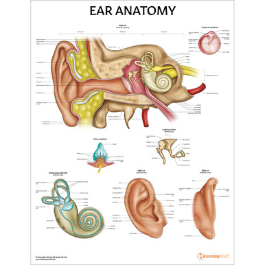
The ear is a complex, shell-like structure responsible for hearing and equilibrium (balance). It can be divided into three anatomical regions; the external, middle and inner ear. Keep reading to learn about the anatomy of this intricate organ, including the process of hearing, its blood supply and innervation.
External ear
The external ear, also known as the outer ear, is the visible part of the hearing apparatus. It consists of the auricle (pinna), the external auditory canal and the lateral surface of the tympanic membrane. It is responsible for capturing and directing sound waves from the external environment onto the tympanic membrane for further transmission.
The auricle is the visible flap of skin on each side of the head. It is composed of skin, elastic cartilage and intrinsic muscles (the three extrinsic muscles are the posterior, superior and anterior auricular muscles, and the six intrinsic muscles are the helicis major and minor, tragicus, anti-tragicus, transverse and oblique muscles). The auricular muscles, however, are now considered to be of little functional significance.
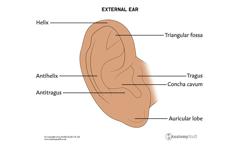
The auricle contains several important anatomical landmarks, such as the helix, tragus and lobule, as shown in the picture below:
The external ear also contains the external auditory canal, sometimes known as the external auditory meatus. This S-shaped tube, which is approximately 2.5cm long, is a pathway from the external ear to the middle ear. One-third of this canal (the part continuous with the auricle) is cartilaginous, and the medial two-thirds are surrounded by bone. The narrowest part is located at the junction of the cartilaginous and bony portions and is known as the isthmus.
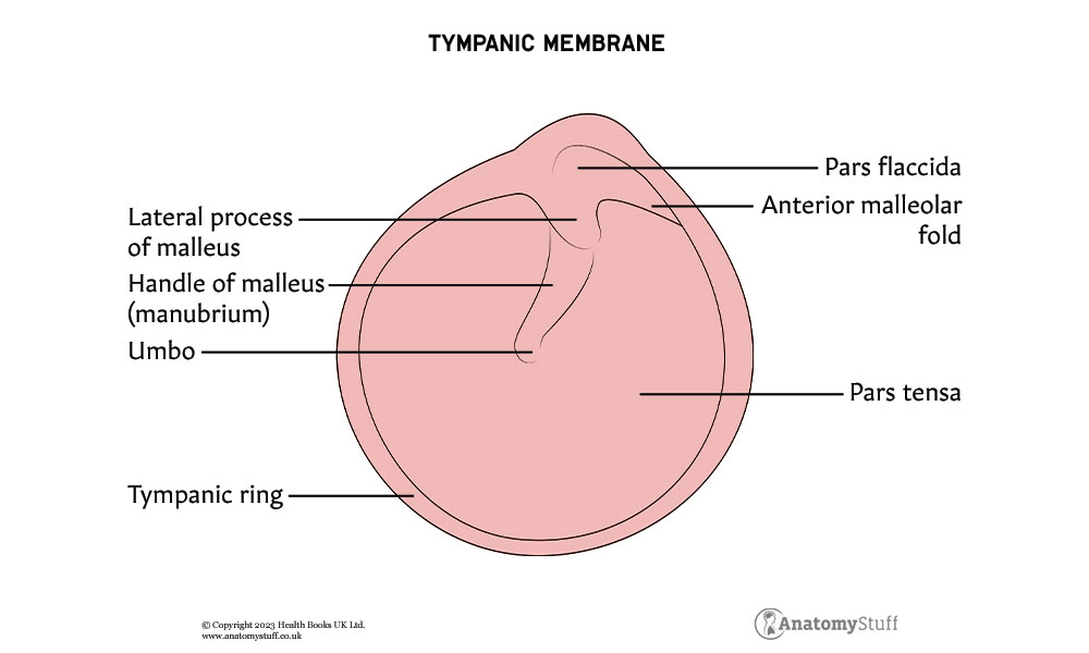
The final section of the external ear is the lateral surface of the tympanic membrane, also known as the eardrum. It lies at the distal end of the external auditory canal, separating the external ear from the middle ear (also known as the tympanic cavity). This thin, oval membrane is comprised of three layers: an outer cutaneous layer, the fibrous middle layer, and a layer of mucous membrane. It can also be divided into two main parts; pars tensa and pars flaccida.
The pars tensa is the tense, main portion of the tympanic membrane with a thickened outer margin which forms a fibrocartilaginous ring called the tympanic ring. The pars flaccida which is also known as the Shrapnell’s membrane, is the flaccid part of the membrane (as its name suggests). It lies just above the lateral process of the malleus. Discover more about this bone in the middle ear section below.
Middle ear
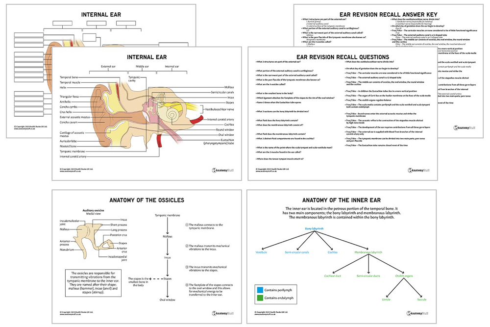
The middle ear or tympanic cavity is a rectangular region located in the petrous portion of the temporal bone. It consists of three bones called ossicles, the oval window, the round window and the Eustachian tube.
The ossicles are responsible for transmitting vibrations from the tympanic membrane to the inner ear. They are named after their shape; malleus (hammer), incus (anvil) and stapes (stirrup). The malleus connects to the tympanic membrane and transmits mechanical vibrations to the incus, which then transmits the vibrations to the stapes. The stapes connect to the oval window, and this allows for mechanical energy to be transferred to the inner ear. Essentially, the ossicles form a small chain across the middle ear.
The oval window is one of two openings into the cochlea from the middle ear. The footplate of the stapes is attached to its rim by the annular ligament, a ring of fibrous soft tissue.
The round window is the second opening into the cochlea. It bulges outward in response to pressure placed on the oval window by the ossicles.
Finally, the Eustachian tube is a fibrocartilaginous channel connecting the middle ear to the nasopharynx. It is approximately 36mm in length and has two important functions:
- Maintaining equal air pressure between the atmosphere and the middle ear.
- Clearing secretions by propelling mucus from the middle ear into the nasopharynx for elimination.
Interestingly, in children, the Eustachian tubes lies in a more horizontal position which impairs drainage and increases their susceptibility to middle ear infections.
The Eustachian tube also remains closed most of the time, preventing pathogens ascending up towards the middle ear. It only opens during times of positive pressure, such as yawning or swallowing or as the result of a special technique called the Valsalva manoeuvre.
Interesting fact
The stapes is the smallest bone in the body!
Inner ear
Just like the middle ear, the inner ear is located in the petrous portion of the temporal bone. It has two main components; the bony labyrinth and membranous labyrinth:
- The bony labyrinth can be divided into three sections: the vestibule, the semicircular canals and the cochlea, which contain a fluid called perilymph.
- The membranous labyrinth, which is located within the bony labyrinth, consists of the cochlear duct, semi-circular ducts and otolith organs (utricle and the saccule). These organs are separated by a fluid called endolymph.
| Components | Structures | Function |
| Bony labyrinth | Vestibule
|
Central chamber containing the otolith organs (saccule and utricle) which are part of the membranous labyrinth. |
| Semicircular canals
|
Three bony tubes (anterior, lateral and posterior) that are responsible for balance. They signal information to the brain about the direction and speed of rotation of the head. | |
| Cochlea | A spiral-shaped bone that acts as the primary structure for the perception of sound. It contains three distinct fluid compartments:
· Scala vestibuli (contains perilymph) · Scala media (contains endolymph) · Scala tympani (contains perilymph) |
|
| Membranous labyrinth | Cochlear duct (scala media) | Situated within the cochlea and contains the organ of Corti, which lies on the basilar membrane. It is composed of sensorineural hair cells (two types- inner and outer) that are stimulated by changes in fluid and vibration. |
| Semicircular ducts | Located within the semi-circular canals | |
| Otolith organs | Two vestibular sacs which are the organs of balance |
As summarised in the table above, the scala vestibuli and scala tympani both contain perilymph, and the scala media contains endolymph. Perilymph is rich in sodium and low in potassium and calcium, whereas endolymph is rich in potassium and low in sodium and calcium. The scala tympani lies within the outer portion of the cochlea, and it is continuous with the scala vestibule (lining the inner portion of the cochlea) at the helicotrema.
Hearing
Hearing is the transduction of sound waves into a neural signal. The complex yet intricate structures of the external, middle and inner ear work together to facilitate this:
• Sound waves enter the external acoustic meatus and strike the tympanic membrane
• The tympanic membrane vibrates in response to the sound wave
• These vibrations are transferred to the middle ear along the ossicular chain
• The footplate of the stapes contacts the oval window
• The vibrations induce pressure waves in the perilymph of the scala tympani and then the scala vestibule
• Waves press against and transmit wave energy to the scala media through the basilar membrane
• The bending of the hairs of the inner hair cells caused by these vibrations activates a neural response in auditory nerve fibres of the vestibulocochlear nerve
![]()
Muscles
There are two muscles found within the ear, and they are both located in the middle region. The tensor tympani muscle attaches to the malleus and the stapedius that attaches to the stapes. They have a protective function as they contract in response to loud noise, therefore inhibiting the vibrations of the ossicles and reducing the transmission of sound to the inner ear. When the stapedius muscle contracts, this is called the acoustic reflex.
Innervation
The inner ear is innervated by the vestibulocochlear nerve (CN VIII), which divides into the vestibular nerve (responsible for balance) and the cochlear nerve (responsible for hearing).
Blood supply
| Region | Arterial supply |
| External ear | Branches of the external carotid artery |
| Middle ear | Maxillary division of the external carotid artery and branches of the internal carotid artery |
| Inner ear | Bony labyrinth – Anterior tympanic branch (from maxillary artery), petrosal branch (from middle meningeal artery) and stylomastoid branch (from posterior auricular artery)
Membranous labyrinth – Labyrinthine artery, a branch of the inferior cerebellar artery |
Embryology
The development of the ear requires contributions from all three germ layers (endoderm, the ectoderm, and the mesoderm). Growth begins around day 22 of gestation and is mostly complete by the 20th week.

Related Products
View All

AnatomyStuff
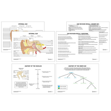
Ear Anatomy Revision Worksheets (Interactive & Printable PDFs)
Now
£5.00
Inc VAT


