


![]()
Introduction
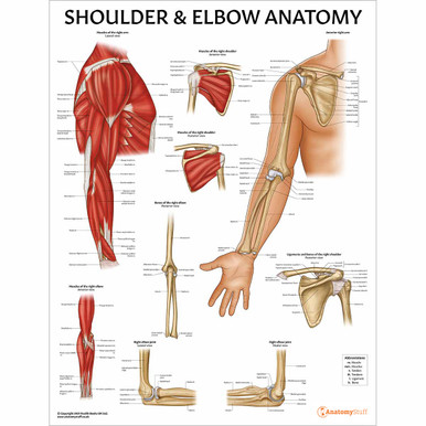
The elbow joint connects the upper arm to the forearm. Specifically, it is the site at which the upper arm’s humerus bone meets the ulnar and radial bones of the forearm. With the help of the surrounding muscles, ligaments and nerves, the elbow joint enables us to bend and straighten our arm. It provides stability to the upper limb and assists us in performing a range of different actions.
Joint Type
The elbow is functionally classified as a hinge-type joint. This means that the movements of the bones at the elbow are in one plane, much like the hinge of the door.
The elbow is structurally classified as a synovial joint and, like all synovial joints, is enclosed by a joint capsule. The inner lining of the joint capsule is composed of the synovial membrane, which secretes synovial fluid. This fluid provides lubrication and allows for smooth movement between the bony surfaces making up the joint.
Osteology
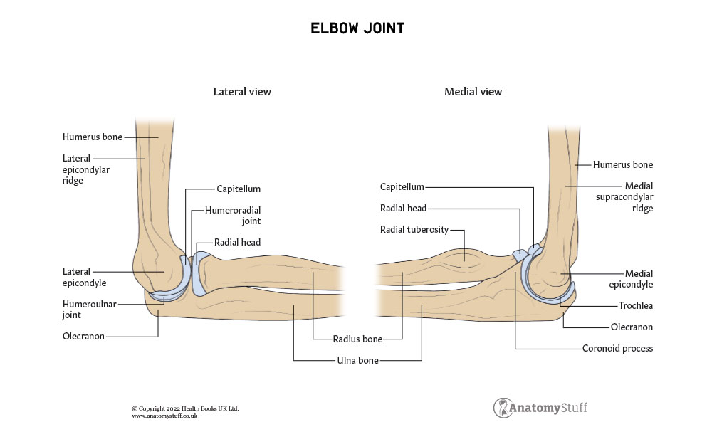
The bones of the elbow are the humerus of the upper arm and the radius and ulna of the forearm. The radius is on the lateral side of the forearm when the body is positioned in an anatomical position. In other words, it is the bone on the side of the thumb finger, and the ulna is the bone on the side of the pinkie finger.
Humerus
This is a long bone of the upper arm and covers the distance from the shoulder to the elbow. The proximal aspect of the humerus articulates with the glenoid fossa of the scapula to form the glenohumeral joint i.e. shoulder joint.
The distal aspect of the humerus articulates the radius and ulna to form the elbow joint, so we will focus on this aspect.
The distal region of the humerus consists of:
• Lateral and medial epicondyles: bony protrusions which can be palpated at the elbow joint. The medial epicondyle is the larger of the two and forms a groove through which the ulnar nerve passes under.
• The trochlea: located at the distal medial articulating surface of the elbow. It extends to the posterior side of the humerus.
• The capitulum: a smooth rounded eminence on the distal anterior articulating surface of the elbow.
• The coronoid, radial and olecranon fossa: depressions found on the distal portion of the humerus
Ulna
The ulna is a long bone of the medial forearm. Proximally, the ulna and humerus articulate, forming part of the elbow joint. Proximally and distally, the ulna articulates with the radius beside it.
The proximal aspect of the ulna consists of:
• Olecranon: a large protrusion of bone which can be palpated at the tip of the elbow
• Coronoid process: a bony ridge projecting anteriorly
• Trochlear notch: a wrench-shaped deep depression formed by the olecranon superiorly and coronoid process inferiorly
• Radial notch: shallow depression on the lateral side of the trochlear notch
• Ulnar tuberosity: ridge distal to a coronoid process where brachialis muscle attaches
Radius
The radius is the long bone of the lateral forearm that lies parallel to the ulna. The proximal aspect of the radius articulates with the humerus and makes up part of the elbow joint. The distal aspect forms the wrist joint. Proximally and distally, the radius articulates with the ulna beside it.
The proximal aspect of the radius consists of:
• Head of the radius: smooth disk-shaped concave surface
• Neck: narrow bone directly under the radial head
• Radial tuberosity: bony protrusion where biceps brachii attaches
Articulating Surfaces
The three bones connect at two separate articulation sites meaning that the elbow consists of two separate joints.
These are:
• Humeroulnar joint: where trochlea of the distal medial humerus and the trochlear notch of proximal ulna articulate
• Humeroradial joint: where the capitulum of the distal lateral humerus and radial head of the proximal radius articulate
The trochlea and capitulum are rounded, smooth convex surfaces which fit tightly into the concave surfaces of the trochlear notch and radial head. This produces the hinge-type features of the elbow joint.
Note that close to the elbow joint is the proximal ulnar joint. This is a separate articulation between the proximal aspect of the ulnar and radial bones. The proximal ulnar joint allows the forearm to rotate.
Ligaments
The elbow is held together and stabilised with the help of 4 different ligaments.
1. Ulnar collateral ligament: a triangular-shaped ligament which can be subdivided into anterior, posterior and inferior parts. It originates from the medial epicondyle of the humerus and extends to the coronoid and olecranon processes of the ulna.
2. Radial collateral ligament: originates from the lateral epicondyle of the ulna and blends with the annular ligament.
3. Annular ligament: a strong ligament forming a ring around the radius head and attaching to the radial notch of the ulna.
4. Quadrate ligament: extends from the inferior border of the radial notch of the ulna and attaches to the neck of the ulna.
![]()
Movement and Musculature
The elbow joint acts like a hinge and is the site where the arm flexes and extends. These movements are performed by the muscles of the upper arm.
Flexion
Flexion is primarily performed by the muscles in the anterior compartment of the arm. These are the biceps brachii, and brachialis Muscles. However, brachioradialis, a muscle of the forearm, is also involved.
Biceps Brachii:
This is a large thick muscle on the anterior side of the upper arm. It is composed of a long head on the lateral side and a short head on the medial side. Both originate from different sites of the scapula; the tendon of the long head from the supraglenoid tubercle and the tendon of the short head from the coracoid process. These heads unite to form the main muscle belly, which inserts into the radial tuberosity of the proximal radius as a single tendon. This tendon continues in the forearm as flattened connective tissue, bicipital aponeurosis.
Brachialis:
The brachialis originates deep to the biceps brachii on the distal anterior part of the humerus. It inserts into the ulnar tuberosity as a single tendon.
The musculocutaneous nerve supplies the biceps brachii, and brachialis muscles.
Brachioradialis:
This muscle originates from the lateral epicondyle of the lateral distal humerus and inserts at the styloid process of the radius. Although primarily located in the forearm, the brachioradialis crosses through the elbow joint and contributes to flexion at the elbow.
Extension
Triceps Brachii
Triceps brachii is a large thick muscle and the only muscle in the posterior compartment of the arm. It is responsible for the extension at the elbow joint.
The triceps brachii consists of three heads; lateral, long, and medial. The lateral head originates at the lateral humerus superior to the radial groove, the long originates at the infraglenoid tubercle of the scapula, and the medial originates at the posterior humerus inferior to the radial groove. The three heads unite as a single tendon with its insertion point onto the olecranon of the ulna.
The radial nerve innervates the brachioradialis muscle and the triceps brachii.
Blood Supply and Innervation
A network of arteries arranged in an anastomotic loop provides blood supply to the elbow. These arteries include the recurrent and lateral branches from the brachial, profunda brachii, ulnar and radial arteries contribute.
Proximal to the elbow joint, the brachial artery bifurcates to the superior and inferior ulnar collateral arteries. The profunda brachii, which runs deep in the arm, branches to the radial collateral and middle collateral arteries. These branches descend down and contribute to the anastomotic loop supplying the elbow joint.
Distally, the radial artery branches to the radial recurrent artery. The ulnar artery divides into the anterior and posterior recurrent arteries. These arteries travel up to the elbow and anastomose with branches of the brachial and profunda brachii arteries.
The anterior part of the elbow is innervated by the medial, musculocutaneous and radial nerves, and the posterior part by the ulnar nerve. The ulnar nerve runs below the skin at the elbow and is responsible for the sensation people get when they hit their ‘funny ‘bone’.

Related Products
View All
AnatomyStuff
Elbow Injuries and Conditions Anatomy Chart / Poster - Laminated
Now
£14.00
Inc VAT

AnatomyStuff
Elbow Anatomy Revision Worksheets (Interactive & Printable PDFs)
Now
£5.00
Inc VAT


