


![]()
Introduction
The eye is a sensory organ that is responsible for detecting visible light. Each one of the two eyeballs is located in the bony orbit, a bilateral cavity in the skull. The orbit also contains extraocular muscles, eyelids, lacrimal glands, blood vessels, nerves and orbital fat. Keep reading to learn all about the anatomy of this complex yet fascinating organ, including its blood supply, innervation and embryological origin.
Layers and compartments
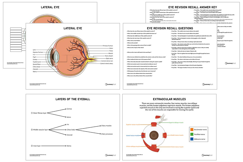
The wall of the eyeball can be divided into three layers:
• An outer fibrous layer consisting of the cornea and sclera
• A middle vascular layer (also known as the uvea) consisting of the iris, ciliary body and choroid
• An inner layer consisting of the retina
The eyeball can also be separated into different compartments:
• Anterior chamber – the space between the cornea and the iris
• Posterior chamber – the space between the iris and the ciliary body
• Vitreous chamber – the largest space behind the lens and in front of the optic nerve
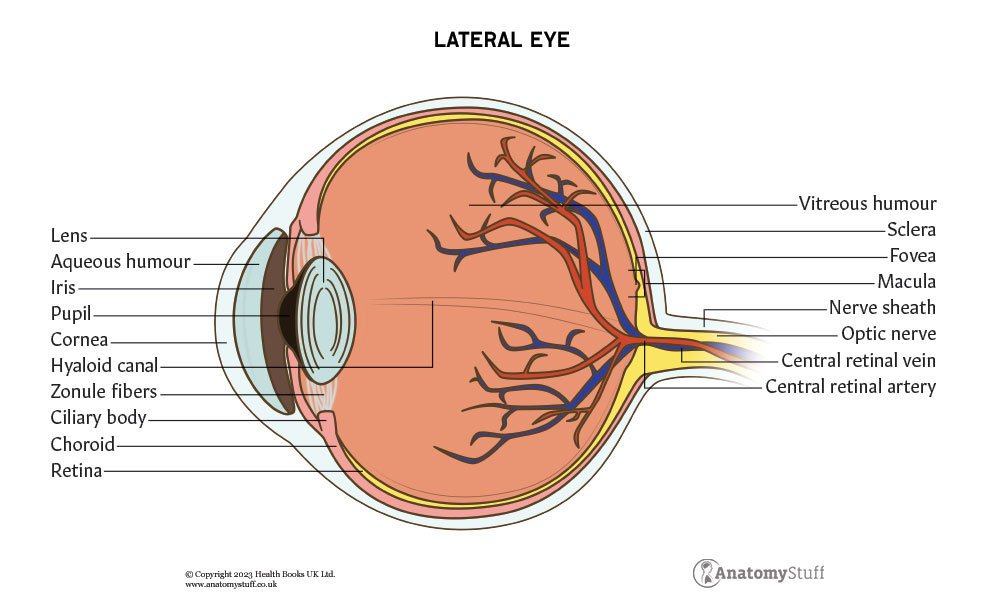
Cornea
The cornea is the outermost, clear layer of the eye, which is located in front of the anterior chamber, iris, and pupil. Its main function is to refract (bend) light, allowing it to pass freely through the pupil and enter the eye. It has five distinct layers:
• Epithelium
• Bowman’s membrane
• Stroma
• Descemet’s membrane
• Endothelium
The cornea borders the sclera at a transitional zone called the corneal limbus. Here, the epithelium gradually becomes thicker.
Sclera
The sclera is also known as the ‘white of the eye’. It comprises dense connective tissue and has four distinct layers: episclera, stroma, lamina fusca, and endothelium. As mentioned above, it is continuous with the cornea at the corneoscleral junction (corneal limbus) and terminates posteriorly at the optic nerve canal. It covers the entire eyeball except for the cornea.
Choroid
The choroid is the posterior portion of the middle layer of the eye. It is a thin and highly vascular connective tissue layer that lines the sclera’s internal surface. Its main function is to provide oxygen and nourishment to the outer retina.
Ciliary body
In the anterior portion of the ‘eye’s middle layer, the choroid becomes the ciliary body. It is composed of ciliary processes and ciliary muscle. The ciliary body has three main roles:
- Accommodation (changing the shape of the lens to focus on near or distant objects)
- Holding the lens in place
- Production of aqueous fluid, a watery fluid found in the anterior and posterior chambers
The lens are suspended by fine ligaments called zonules, which are also attached to the ciliary body. When the ciliary muscle contracts, the lens becomes more rounded. When the ciliary muscle relaxes, the lens transforms into a flatter shape. This is the process of accommodation.
Iris
The iris is a pigmented, circular muscle located between the cornea and the lens. It has an opening in its centre called the pupil. The iris regulates how much light passes through the pupil and enters the eye.
Interesting fact!
The concentration of melanin in the iris determines the eye colour.
Retina
The third and innermost layer of the eyeball is the retina. It contains many layers, including:
• Pigmented layer
• Neural layer
• The neural layer
The neural layer contains three main cells types:
• Photoreceptor cells
• Bipolar cells
• Retinal ganglion cells
Photoreceptors are specialised cells that convert light rays into nerve impulses. This process is called phototransduction. There are two types of photoreceptor cells – rods and cones:
| Type of photoreceptor cell | Amount (approximately) | Location | Function |
| Rods | 100 million | Outside the fovea | Allow us to see in dim light |
| Cones | 6 million | Concentrated in the fovea (apart from the centre of it) | Allow us to see in colour vision (results from the stimulation of different combinations of blue, green, and red cones) and responsible for visual acuity |
The fovea is a small depression in the centre of the retina.
Rods are much more sensitive to light than cones. Hence, they are mainly responsible for vision during times of low lighting.
Eventually, the visual information from these photoceptor cells reaches the optic disc. This area is also called the blind spot because it contains no rod or cone cells and is where nerve fibres and blood vessels pass through the eyeball.
![]()
Lens
The lens is suspended behind the pupil and iris. It is encircled by the ciliary processes and is attached to them by the zonular fibres. As mentioned above, the ciliary muscles can alter the lens’s shape, which is known as accommodation. The lens also refracts light, focusing it onto the retina.
The eyeball can be separated into three compartments; the anterior, posterior and vitreous chambers. It is the lens that divides the eyeball into the anterior cavity and the vitreous cavity. The anterior cavity is then further divided into the anterior and posterior chambers.
Aqueous and vitreous humour
The anterior and posterior chambers are filled with aqueous humour, a watery fluid that nourishes the cornea and lens and maintains ocular pressure (eye pressure). It is produced by the ciliary epithelium lining the ciliary processes in the posterior chamber and then flows through the pupil into the anterior chamber.
The majority of aqueous fluid from the anterior chamber is drained through the trabecular meshwork (a group of specialised cells) into the canal of Schlemm, which drains to episcleral veins. The remainder of aqueous humour exits through a different pathway called the uveoscleral route.
Vitreous humour is a clear, gel-like substance located within the vitreous cavity. It is comprised almost entirely of water and does not undergo rapid replacement, unlike the aqueous humour.
Extraocular muscles
There are seven extraocular muscles; four rectus muscles, two oblique muscles, and the levator palpebrae superioris muscle.
The levator palpebrae superioris muscle is the only one involved in raising the superior eyelid. The rest of the muscles are responsible for moving the eyeball, and their exact function is listed in the table below:
| Muscles | Main function | Innervation |
| Superior Rectus | Elevation of the eyeball | Oculomotor nerve |
| Inferior Rectus | Depression of the eyeball | Oculomotor nerve |
| Medial Rectus | Adducts the eyeball | Oculomotor nerve |
| Lateral Rectus | Abducts the eyeball | Abducens nerve |
| Superior Oblique | Depresses, abducts and medially rotates the eyeball | Trochlear nerve |
| Inferior Oblique | Elevates, abducts and laterally rotates the eyeball | Oculomotor nerve |
Eyelids
The upper and lower eyelids are soft tissue structures which cover the base of the orbits, protecting the eye from injury.
Lacrimal gland
The lacrimal gland is also known as the tear gland. It is located above the eyeball in the bony orbit and produces tears to keep the surface of the eyeball lubricated.
Conjunctiva
The conjunctiva is a thin mucous membrane that allows the eyelid to move freely over the eye. It lines both the inner surface of the eyelids and the sclera.
Bony orbit
The bony orbits are symmetrical cavities within the head. They contain the eyeballs, extraocular muscles, eyelids, nerves and blood vessels. The rest of the cavity is filled with orbital fat.
Blood supply
The main blood supply of the eye arises from the ophthalmic artery, the first branch of the internal carotid artery. It gives off several important branches, including the central retinal artery, which supplies the inner layers of the retina, the lacrimal artery, the eyelids, the lacrimal gland, and the conjunctiva.
Venous drainage corresponds with the arterial supply; ophthalmic veins and a central retinal vein drain blood from the eye.
Innervation
Overall, six cranial nerves innervate different structures in the eyes:
| Cranial nerve | Function |
| Optic nerve | 1) Senses the incoming light and image displayed on the retina and then transmits this information to the cerebral cortex
2) Works with the oculomotor nerve to change pupil size |
| Oculomotor nerve | 1) Innervating the major of the extraocular muscles of the eyes (see table above)
2) Works with the optic nerve to change pupil size |
| Trochlear nerve | 1) Motor innervation to the superior oblique muscle only |
| Trigeminal nerve | 1) Afferent part of the corneal and lacrimation reflex |
| Abducens nerve | 1) Motor innervation to the lateral rectus muscle only |
| Facial nerve | 1) Motor innervation of the orbicularis oculi muscle (responsible for eye closure and blinking)
2) Efferent part of the corneal and lacrimation reflex |
To change pupil size, the optic nerve conducts the afferent impulse to the brain. Then the oculomotor nerve will constrict the pupils if the light is bright, allowing pupil dilation when the light is dim.
Embryology
Eye development begins in the third to fourth week of gestation. Ocular structures have a neuroectodermal origin.

Related Products
View All


AnatomyStuff
Eye Anatomy Revision Worksheets (Interactive & Printable PDFs)
Now
£5.00
Inc VAT


