


![]()
Introduction
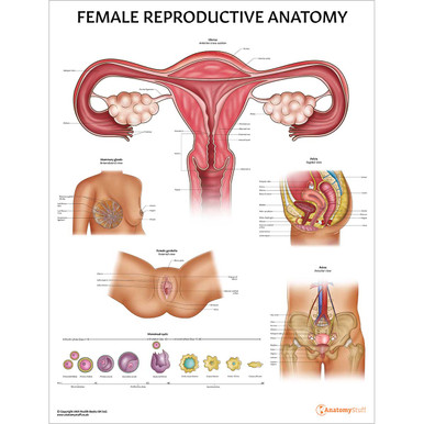
Breasts are organs made up of fat, mammary glands and connective tissues and ligaments present in both males and females. However, only female breasts are well developed and therefore play a role in the reproduction process. Female Breasts contain mammary glands. These are modified sweat glands that are necessary for lactation.
• The female breasts extend from the 2nd rib to the 6th rib along the mid-clavicular line.
• Roughly two-thirds of the breast rests on the pectoral fascia that covers the pectoralis major. The other third rests on the fascia covering the serratus anterior muscle.
• During puberty, female breasts grow due to the deposition of fat in this area and due to glandular development.
• The nipple is at the breast’s centre and composed of smooth muscle fibres. It is surrounded by pigmented skin known as areole. During pregnancy, glands in the areolae secrete an oily lubricant to help protect the nipple.
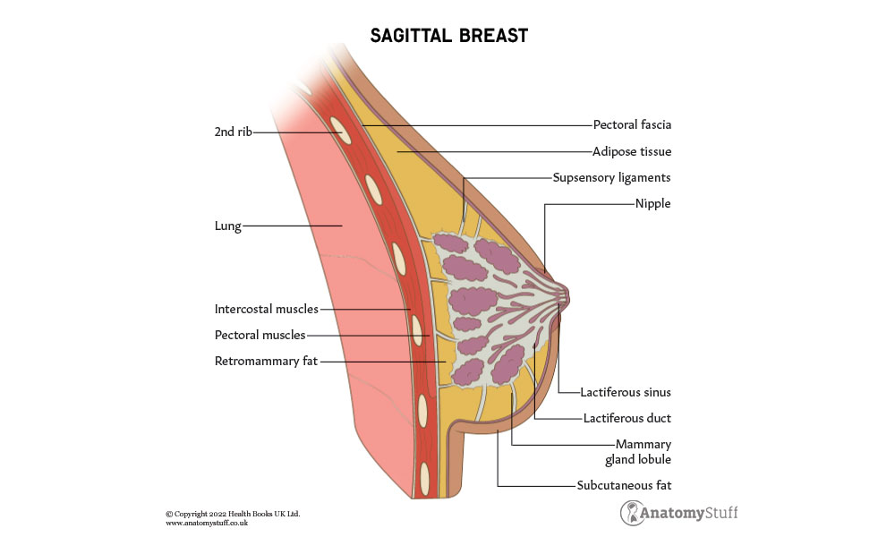
Lobules and Ducts
The mammary gland consists of ducts and secretory lobules. These structures converge and open around the nipple.
Lactiferous ducts
• Each breast is divided into 15-25 lobes by fibrous septa
• These lobes drain into ten major lactiferous ducts
• Lobes further divide into lobules, with each lobule containing 10-100 alveoli
• These ducts open on the surface of the nipple
• A large lactiferous duct leaves each lobe and widens to form a lactiferous sinus
Alveoli
• A collection of alveoli make up a lobule
• Alveoli are sites of milk synthesis and storage in lactating women
• Epithelial lactocytes lining the alveoli produce milk
• Myoepithelial cells surround alveoli and contribute to a dense network of capillaries. Contraction of myoepithelial cells squeezes the alveoli and allows for the ejection of milk into the larger duct until it eventually reaches the nipple
Lobuloalveolar ducts
• Milk from each alveoli is collected by the lobuloalveolar ducts, which join together to form a single lactiferous duct.
Ligaments and Connective Tissue of the Breast
Nipple
• The nipple contains longitudinal and horizontal smooth muscle fibres
• It has a sphincter-like function
• Nipple ducts are usually 0.5mm in diameter
Areola
• This is a circular area of pigmented skin that surrounds the nipple
• The lactiferous sinus is present just deep to the areola
Cooper’s ligaments
• The mammary glands are attached to the overlying skin through suspensory ligaments called Cooper’s ligament
• These ligaments are continuous with the skin’s dermis and support the breast. They also separate the secretory lobules of the breast
Chassaignac bursa
• Also known as the retro mammary bursa, this is a space that contains loose connective tissue and is present between the breast and posteriorly the deep layer of fascia covering the pectoral muscles
• This space help in the mobility of the breast over the thoracic wall and is useful in reconstructive plastic surgery
![]()
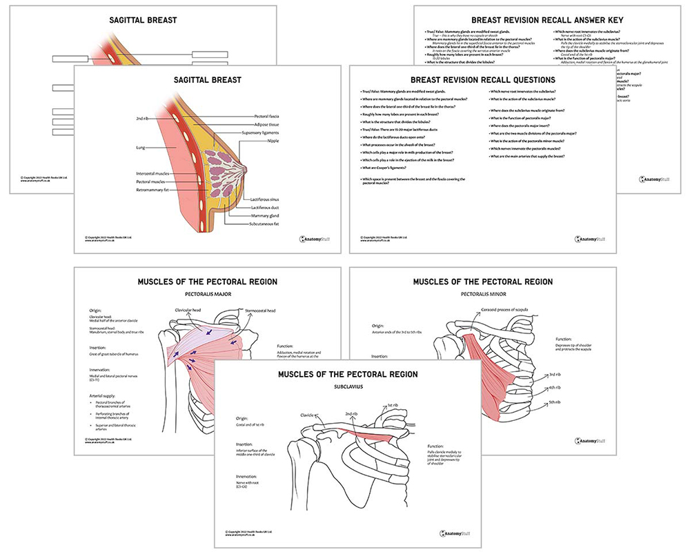
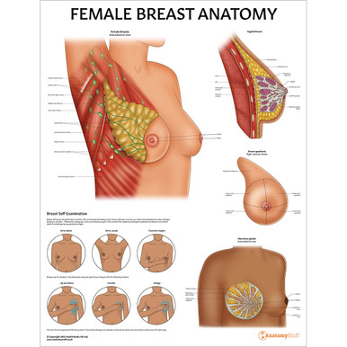
Muscles of the Pectoral Region
A number of muscles in the pectoral region originate at the anterior thoracic wall and insert at different bony features of the upper limbs. These muscles help stabilise and provide movement to the axio-appendicular region. These muscles are the pectoralis major, pectoralis minor and subclavius.
Arterial Supply of the Breasts
Vessels of the thoracic wall are involved in the supply and drainage of the breast. This means that most vessels relating to the breast also supply and drain other structures in the thoracic wall. The breast receives arterial supply from many sources.
Subclavian artery – medial mammary branches and anterior intercostal branches of the internal thoracic artery, which originate from the subclavian artery
Axillary artery – lateral mammary branches and thoracoacromial arteries, which are branches of the axillary artery
Thoracic aorta – specifically the posterior intercostal arteries which run in intercostal spaces and are branches of the thoracic aorta
Venous Drainage of the Breast
The pathway of the veins in the breast is parallel to that of the arteries. All veins ultimately drain into:
• The axillary veins
• Internal thoracic vein
• Intercostal veins
Lymphatic Drainage of the Breast
Lymphatic drainage of the breast is clinically very relevant as when cancer of the breast metastasis, it spreads through this route. Lymph fluid in the breasts, originating in the lobules, nipples, and areola, travels to the subareolar lymphatic plexus. This lymph is then drained in different ways. Lymph from axillary nodes ultimately drains into the subclavian lymphatic trunk, whereas lymph from parasternal nodes ultimately drains into the thoracic or right lymphatic duct.
Axillary lymph nodes – most of the lymph from the breast drains into axillary lymph nodes
Pectoral (anterior) nodes – before draining into the axillary nodes, most lymph drains into the pectoral nodes; however, some may drain directly to other axillary nodes or to interpectoral, deltopectoral, supraclavicular or inferior deep cervical nodes
Parasternal lymph nodes – some lymph from the medial breast quadrants drains into the parasternal lymph nodes
Abdominal lymoh nodes – these mostly collect lymph from inferior breast quadrants.
Nerve Supply
The breast is supplied by the anterior and lateral cutaneous branches of the 4-6th intercostal nerves, which contain both sensory and autonomic functions.
Milk production and secretion are not controlled by these nerves but by the hormones prolactin and oxytocin from the pituitary gland.

Related Products
View All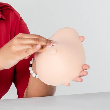

AnatomyStuff
Female Breast Anatomy Revision Worksheets (Interactive & Printable PDFs)
Now
£5.00
Inc VAT


