


![]()
Introduction
The average human will have walked 110,000 miles in their lifetime, and this is the equivalent of walking 5 times around the earth! Humans are only able to do this with the special and unique design of the foot. Our feet not only enable us to stand upright, walk, run and jump, but they also carry the weight of our entire body. The ankle joint, anatomically referred to as the talocrural joint, enables our feet to perform a range of different movements with the help of numerous bones, ligaments, muscles and tendons.
Osteology
The rigid bony framework of the foot provides mechanical support to the surrounding soft tissues and enables the weight-bearing function. There are a total of 26 bones in the foot, and these are divided into 7 tarsals, 5 metatarsals, and 14 phalanges.
Tarsals
The tarsals are located most proximally in the foot and are a cluster of 7 irregularly-shaped bones. They are arranged into a proximal, intermediate and distal rows. The proximal row consists of the talus superiorly and the calcaneus inferiorly. The intermediate row only consists of the navicular bone, and the distal row consists of the cuboid and three cuneiforms – lateral, intermediate and medial.
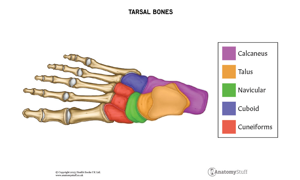
Talus
The talus is the most superior bone of the foot and connects the foot to the rest of the body via the talocrural joint. In doing so, it transmits the weight of the body from the tibia of the lower leg to the calcaneus, i.e. the heel. The talus articulates superiorly with the tibia and fibula of the lower leg forming the talocrural joint. Inferiorly it articulates with the calcaneus forming the subtalar joint, and anteriorly with the navicular forming the talonavicular joint.
Calcaneus
The calcaneus, colloquially referred to as the heel, is the weight-bearing and largest bone of the foot. It articulates with the talus superiorly via the subtalar joint and anteriorly to the cuboid via the calcaneocuboid joint. The calcaneal tuberosity is a protrusion on the posterior aspect of the foot where the Achilles tendon attaches
Navicular
Navicular means boat-shaped, and this bone is located on the medial side of the foot. It articulates with the talus posteriorly at the talonavicular joint, cuboid laterally and cuneiform bones anteriorly. The tibialis posterior tendon attaches to a small tuberosity on the plantar surface, i.e. sole of the navicular.
Cuboid
The cuboid is located anterior and most lateral in the distal row of the foot. It articulates to the calcaneus posterior to it via the calcaneocuboid joint.
Cuneiforms
The three cuneiforms (lateral, intermediate and medial) articulate with the navicular posteriorly and metatarsals anteriorly. They are shaped like a wedge, and this forms the transverse arch of the foot. The tibialis anterior, posterior and fibularis longus attach to the medial cuneiform. The flexor hallucis brevis attaches to the lateral cuneiform.
Metatarsals
There are 5 metatarsal bones, with 1st being the most medial and 5th being the most lateral. Each metatarsal consists of a head, neck, shaft and base, with the head being the most distal. They articulate proximally with the tarsal bones at the tarsometatarsal joint, laterally with each other at the intermetatarsal joints and distally with the phalanges at the metatarsophalangeal joints.
Phalanges
The phalanges are the bones of the toes, and there are a total of 14 of them. Each phalanx is separated into a base, shaft and head like the metatarsals. The big toe has proximal and distal phalanges, whereas the rest of the digits each have proximal, middle and distal phalanges.
Joint Type
Talocrural Joint
The talocrural joint is the complex articulation between the tibia and fibula of the lower leg and the talus of the foot. The tibia is the larger of the two bones and forms the medial border of the talocrural joint. The fibula forms the lateral border. The tibia, fibula and talus articulate at 3 sites:
- The medial malleolus, a bony projection on the distal end of the tibia, articulates with the medial surface of the talus.
- The lateral malleolus on the distal end of the fibula articulates with the lateral surface of the talus.
- The distal end of the tibia articulates with the trochlea of the talus, which is a superior rounded surface on the talus
The talocrural joint is known as a mortise joint. This is because the malleoli of the tibia and fibula connect via strong tibiofibular ligaments forming a bracket-shaped socket, the mortise. The body of the talus is shaped like a wedge and snugly fits into the mortise. The talus has a broad anterior aspect making the joint more stable in dorsiflexion, and a narrow posterior aspect making it less stable in plantarflexion.
The talocrural joint is functionally a hinge-type joint, meaning that movement exists in the vertical plane. Therefore, the talocrural joint is responsible for the dorsiflexion (cocking the foot up) and plantarflexion (pressing the foot down) of the foot.
The talocrural joint is structurally classified as a synovial joint. This is because it is surrounded by an articular capsule which is lined by an inner synovial membrane. This membrane produces synovial fluid, which lubricates the joint. The fibres of the articular capsule are weak and thin anteriorly and posteriorly. However, the articular capsule is strengthened medially and laterally by collateral ligaments. The synovial membrane of the joint capsule projects superiorly between the tibia and fibula up to the interosseus tibiofibular ligament.
![]()
Subtalar Joint
Whilst the talocrural joint, i.e. the true ankle joint, is responsible for dorsiflexion and plantar flexion of the foot, the subtalar joint between the talus and calcaneus facilitates for inversion and eversion of the foot. It is assisted by the talonavicular and calcaneocuboid joints, which, combined together, are known as transverse tarsal joints. The tarsal bones are organised in such a way that the talus and calcaneus move via these joints, whilst the navicular and cuboid bones are fixed.
Interphalangeal Joints
The big toe consists of two phalanges and two joints- the metatarsophalangeal joint at the base of the toe and the interphalangeal joint between the two phalanges. The rest of the digits consist of three phalanges and three joints; the metatarsophalangeal joint at the base, the proximal interphalangeal joint in the middle of the digit and the distal phalangeal joint closest to the tip of the digit.
Ligaments
The ligaments of the foot reinforce the bony framework and stabilise the joints enabling weight-bearing function. Moreover, they support the arches of the foot, and this provides the foot with flexibility so that it can absorb shock during movement.
Ligaments within the Foot
Plantar Fascia
The plantar fascia is a very strong and thick ligament on the sole of the foot that connects the heel to the front of the foot. Its main role is to absorb shock whilst running, walking or jumping. It originates from the medial calcaneal tubercle of the calcaneus and inserts to the base of the five metatarsal bones.
Plantar Calcneonavciular Ligament Complex
Also referred to as the spring ligament complex, this is a set of three ligaments located on the underside of the foot. They provide support to the medial longitudinal arch of the foot and also support the talus head during weight-bearing activity.
Calcaneocuboid Ligaments
These are a group of four ligaments which support the medial and lateral longitudinal arches and stabilise the calcaneocuboid joint. They consist of short plantar and long plantar ligaments and dorsolateral and medial calcaneocuboid ligaments.
Lisfranc Ligament
This is a strong ligament which connects the tarsals to the metatarsals and provides stability to the midfoot. Specifically, its origin site is from the lateral aspect of the medial cuneiform, and the insertion site is at the medial aspect of the base of the 2nd metatarsal.
Ligaments of the Ankle Joint
Medial Ligaments
The deltoid ligament complex is a group of four ligaments arranged in a triangle formation. Their primary role is to prevent over-eversion of the foot. They fan out from the medial malleolus of the tibia and attach to the talus, calcaneus and navicular.
Lateral Ligaments
The main role of the three separate lateral ligaments is to prevent over-inversion of the foot. Whilst all three ligaments originate from the lateral malleolus of the fibula, they insert at different sites. The anterior talofibular ligament inserts to the anterior aspect of the talus. It is the weakest of the three and is a common site for injury. The posterior talofibular ligament inserts to the posterior aspect of the talus, and the calcaneofibular ligament inserts to the calcaneus.
Musculature and Movement
The foot muscles can be divided into an extrinsic and intrinsic group.
Extrinsic Muscles
The extrinsic muscles originate outside of the foot, from the anterior, posterior and lateral compartments of the leg. They act on the talocrural joint to produce plantarflexion and dorsiflexion and also the subtalar (and intertarsal) joints to produce eversion and inversion.
Intrinsic Muscles
The intrinsic muscles of the foot originate from within the foot and are responsible for the fine motor movement of the foot, i.e. flexion and extension, abduction and adduction of digits.
The intrinsic muscles of the foot can be divided into those located in the dorsal aspect of the foot and those in the plantar aspect of the foot, e.g. sole of the foot.
Dorsal Aspect
The dorsum of the foot is the attachment site for many extrinsic foot muscles. However, there are only two muscles intrinsic to the foot, and these are the extensor digitorum brevis and extensor hallucis brevis. The extensor digitorum brevis lies deep to the tendon of the extensor digitorum longus, whereas the extensor hallucis brevis lies medial to it.
| Origin | Insertion | Action | |
| Extensor digitorum brevis | calcaneus, interosseous talocalcaneal ligament and inferior extensor retinaculum | proximal phalanx of big toe and extensor tendons of 2nd to 4th digits | assist in extension of 1st to 4th digits at metatarsophalangeal and interphalangeal joints, alongside extensor digitorum longus. |
| Extensor hallucis brevis | calcaneus, interosseous talocalcaneal ligament and inferior extensor retinaculum | proximal phalanx of big toe | assists extensor hallucis longus in extension of big toe at its metatarsophalangeal joint |
Plantar Aspect
There are 10 intrinsic muscles in the plantar aspect of the foot, and these can be subdivided into four layers, the first layer being most superficial to the sole of the foot.
The first layer consists of three muscles. The abductor hallucis is on the medial side of the sole and contributes to the soft tissue bulge on the sole. The flexor digitorum brevis is located at the centre of the sole, and the abductor digiti minimi is on the lateral side.
The second layer consists of two muscle groups. The quadratus plantae is separated from the first layer by lateral plantar nerves and vessels. There are also four lumbrical muscles in the second layer of the plantar aspect of the foot.
The third layer consists of three muscles. The flexor hallucis brevis is located on the medial side of the foot, and the flexor digiti minimi brevis is located on the lateral side. The adductor hallucis is in-between and has two heads.
The fourth most deep layer of the plantar aspect of the foot consists of three plantar and four dorsal interossei, both of which are situated between the metatarsals. Each of the plantar interossei originates from a single metatarsal, whereas each of the dorsal interossei originates from two metatarsals.
| First layer | Origin | Insertion | Action |
| Abductor hallucis | medial tubercle of the calcaneus, the flexor retinaculum and the plantar aponeurosis
|
base of the proximal phalanx of the big toe | flexes and abducts big toe |
| Flexor digitorum brevis | medial tubercle of the calcaneus and the plantar aponeurosis | middle phalanges of 2nd – 5th digits
|
flexion of 2nd-5th digits at proximal interphalangeal joints |
| Abductor digiti minimi | medial and lateral tubercles of calcaneus and plantar aponeurosis | lateral base of proximal phalanx of 5th digit
|
flexion and abduction of 5th digit |
|
Second layer |
|||
| Qudratus plantae | medial and lateral plantar surface of the calcaneus | flexor digitorum longus tendons
|
flexes 2nd – 5th digits alongside flexor digitorum longus |
| Lumbricals | Each lumbrical spans medially from its respective tendon of the flexor digitorum longus
|
each lumbrical inserts to the extensor hood of one of the 2nd – 5th digits | flexion of metatarsophalangeal joints, and extension of interphalangeal joints |
|
Third Layer |
|||
| Flexor hallucis brevis | plantar surface of lateral cuneiforms and cuboid, tendon of posterior tibialis tendon
|
base of proximal phalanx of big toe | flexes proximal phalanx of big toe at metatarsophalangeal joint |
| Adductor hallucis | Transverse head from plantar ligaments of metatarsophalangeal joints, oblique head from bases of 2-4th metatarsals | Both heads to base of proximal phalanx of big toe | Adduct big toe and help in formation of transverse arch of foot |
| Flexor digiti minimi brevis | Base of 5th metatarsal | Proximal phalanx of 5th digit | Flexes proximal phalanx of 5th digit |
|
Fourth layer |
|||
| Plantar interossei | All 3 from medial side of 3rd– 5th metatarsals | Medial side of phalanges of 3rd -5th digits | Adducts and flexes 3rd to 5th digits at metatarsophalangeal joints |
| Dorsal interossei | All 4 from sides of 1-5th metatarsals | Medial/ lateral side of proximal phalanx of 2nd– 4th digits | Abduct and flexes 2nd -4th digits at metatarsophalangeal joints |
Neurovascular Supply
Arterial Supply
The arterial supply to the ankle joint is provided by the malleolar branches of the posterior tibial and fibular arteries as well as the anterior tibial artery. They form an anastomoses around the malleoli.
The foot itself is supplied by the dorsalis pedis artery and the posterior tibial artery.
• The dorsalis pedis artery travels over the tarsal bones before heading inferiorly to the sole of the foot. It is the continuation of the anterior tibial artery and joints with the lateral plantar artery to form the deep plantar arch. It supplies the tarsals, the dorsal part of the metatarsals and partially supplies the digits.
• The posterior tibial artery travels to the sole of the foot via the tarsal tunnel, where it bifurcates to lateral and medial plantar arteries. These arteries provide blood supply to the plantar aspect of the foot and also the toes via the deep plantar arch.
Note that the tarsal tunnel is a passageway for tendons, nerves and vessels to enter the foot from the posterior leg. It is formed by the tibia, talus and calcaneus inferiorly and connective tissue superiorly. The posterior artery and vein and the tibial nerve travel through the tarsal tunnel.
Dorsal Venous Drainage
The dorsal venous arch is mainly responsible for the venous drainage of the foot. From the arch, the blood is drained to superficial veins, some of which travel deep in leg to form the anterior tibial vein.
The medial and plantar veins from the plantar aspect of the foot join to form posterior tibial and fibular veins. These veins travel alongside their corresponding arteries to drain the foot and ankle joints. They then travel up the leg, and at the posterior aspect of the knee, the anterior tibial, posterior tibial and fibular veins combine to form the popliteal vein. The popliteal vein enters the thigh via the adductor canal.
Nerve Supply
The branches of the tibial, superficial fibular and deep fibular nerve innervate the ankle joint and foot. These branches are derived from the roots of L4-S2.

Related Products
View All

AnatomyStuff
Foot & Ankle Anatomy Revision Worksheets (Interactive & Printable PDFs)
Now
£5.00
Inc VAT


