


Introduction
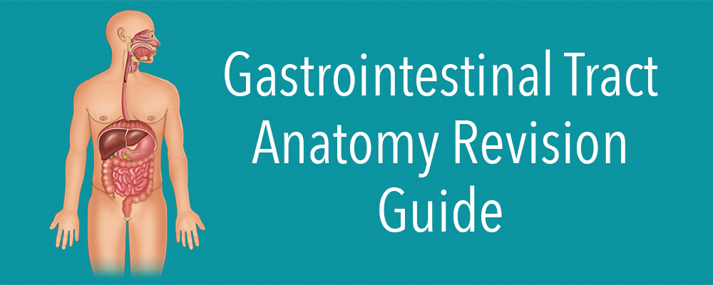
The gastrointestinal (GI) tract, also known as the alimentary tract, is a continuous channel throughout the body which facilitates the transportation, digestion and absorption of food. It consists of the oral cavity, pharynx, oesophagus, stomach, small intestine and large intestine. Keep reading our Gastrointestinal Tract Anatomy Revision guide to learn more about this important system, including its accessory organs, blood supply, innervation and embryological origin. Pair it with our Gastrointestinal Tract Anatomy Revision Worksheets for the ultimate revision combo.
Overview
The processes of digestion can be divided into six steps:
• Ingestion
• Propulsion
• Mechanical digestion
• Chemical digestion
• Absorption
• Defecation
Ingestion involves the entry of food through the mouth. The act of chewing crushes and grinds the food down, increasing its surface area. This process is called mastication. In the mouth, the food also mixes with saliva, which aids with digestion.
The food is then propelled into the oesophagus by the act of swallowing. Throughout the GI tract, it is propelled again by an involuntary process called peristalsis (more information about this is below). Both swallowing and peristalsis are examples of propulsion.
Mechanical digestion is the process of making the food smaller to increase both surface area and mobility. It includes mastication, tongue movements and segmentation (discussed more below). The semifluid mass of partly digested food is called chyme.
Digestive enzymes break Food down from complex molecules into smaller chemical building blocks. This process takes place in the small intestine and is called chemical digestion.
Absorption is when the products of chemical digestion cross the mucosa and enter the lymph or the bloodstream. Like chemical digestion, it primarily takes place within the small intestine.
Defecation is the process of eliminating undigested materials from the body.
Histology
Most sections of the GI tract are composed of four layers of tissue (from deep to superficial):
• Mucosa
• Submucosa
• Muscularis propria
• Serosa or adventitia
The mucosa, which comes into direct contact with chyme, is made up of three layers:
• Epithelium – where most of the digestion, absorption and secretion occurs
• Lamina propria – a layer of connective tissue
• Muscularis mucosae – a very thin double-layer of smooth muscle
The submucosa is a layer of loose connective tissue containing larger blood vessels, nerves, mucous-secreting glands and nerves.
The muscularis propria consists of two muscle layers – the inner circular layer and the outer longitudinal layer (in the stomach, there is a third oblique layer). Between the inner layer and the outer layer lies the myenteric plexus, which facilitates peristalsis to occur. Additionally, the thickness of this muscular layer differs along the GI tract. For example, it is thicker in areas like the large intestine and thinner in the small intestine.
Finally, the outermost layer of the GI tract is comprised of either serosa or adventitia. Serosa covers intraperitoneal structures, and adventitia covers retroperitoneal structures. They both contain blood vessels, nerves and lymphatics.
Movements
Two types of movements occur in the gastrointestinal (GI) tract; peristalsis and segmentation. Peristalsis is the involuntary contraction and relaxation of longitudinal and circular muscles. This allows for the propulsion of contents throughout the GI tract.
Segmentation describes localised contractions of circular muscle. It mainly occurs in the small intestine and moves the contents back and forth rather than propelling them forward, like peristalsis. By moving food back and forth in the intestinal lumen, segmentation mixes food with digestive juices and facilitates absorption.
Oral cavity
The GI tract begins in the oral cavity, which consists of the lips, tongue, palate and teeth. These structures work together to facilitate the intake and digestion of food and water, as well as several other life-sustaining processes, such as the formation of speech and respiration.
Most adults have 32 teeth. They can be divided into 4 subcategories:
• Incisors – designed for cutting
• Canines – designed for tearing
• Premolars – designed for grinding
• Molars – designed for grinding
The tongue consists of intrinsic and extrinsic muscles. It is also populated with taste buds that facilitate gustatory sensation.
You can read more about it in our Tongue Anatomy Revision Guide.
Pharynx
The pharynx is a fibromuscular tube located in the midline of the neck. It connects the mouth to the oesophagus, facilitating the passage of solids and liquids. You can discover more about the anatomy of this important organ using our Pharynx Anatomy Revision Guide.
Oesophagus
The oesophagus is a fibromuscular tube connecting the pharynx to the stomach. It lies posterior to the trachea and is roughly 25cm long. It is roughly Its primary function is to allow for the passage of liquids and solids.
It can be divided into three anatomical segments:
• Cervical
• Thoracic
• Abdominal
The cervical segment begins at the cricopharyngeus (one of the pharynx’s inferior constrictor muscles) at vertebrae level C6. It extends to the suprasternal notch, a large depression on the top of the sternum.
The thoracic segment runs from the suprasternal notch to the diaphragm. It pierces the diaphragm at the tenth thoracic vertebra (T10), and this opening is known as the oesophageal hiatus. The right and left vagus nerves and oesophageal branches of the left gastric artery and vein also pass through this opening.
The remaining 2.5cm of the oesophagus is known as the abdominal segment. This section extends from the diaphragm to the fundus of the stomach. It descends and passes through the right crus of the diaphragm, which is one of two tendinous structures arising from the vertebrae.
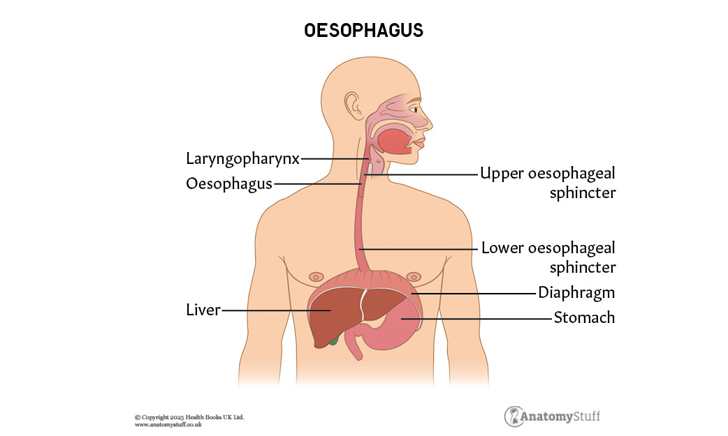
There are two sphincters located in the oesophagus; the upper oesophageal sphincter (UES) and the lower oesophageal sphincter (LES).
The UES is a high-pressure zone located between the pharynx and cervical oesophagus. It primarily comprises the cricopharyngeus muscle and normally remains closed in a contracted position. During swallowing, the muscles temporarily relax, allowing liquids and solids to enter the oesophagus.
The LES is also called the cardiac sphincter. It also remains closed until it involuntarily opens during oesophageal peristalsis. This allows food to enter the stomach.
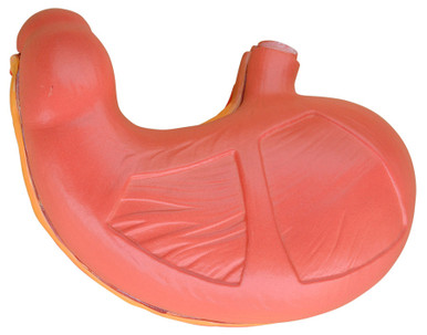
Stomach
The stomach, which is located in the left upper quadrant of the abdomen, is continuous, with the oesophagus proximally and the duodenum distally. This hollow J-shaped organ has many important functions, such as:
• Bulk storage of undigested food
• Mechanical breakdown of food
• Production of important acids, enzymes and hormones
Read more about the stomach by checking out our Stomach Revision Guide.
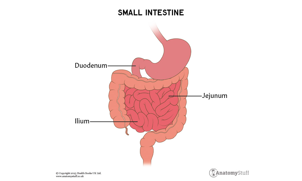
Small intestine
The small intestine lies between the stomach and the large intestine. It is the longest part of the digestive system and measures around 5 metres long. It can be divided into three regions:
• Duodenum
• Jejunum
• Ileum
The duodenum is the shortest section and measures around 20cm in length. It is the only part of the intestine connected to the stomach. It also connects to the jejunum at the ligament of Treitz where (often called the duodenojejunal flexure). It surrounds the pancreas in the shape of a “C” and receives chyme from the stomach, pancreatic enzymes, and bile from the liver. Most chemical digestion takes place here.
There are special mucin-secreting glands called Brunner’ ‘s glands located in the submucosa of the duodenum. They secrete an alkaline fluid containing mucin, protecting the mucosa from the acidic stomach contents.
The jejunum is the middle part of the small intestine and is roughly 2.5 metres long. Its main role is to ensure that the molecules from chemical digestion (primarily in the duodenum) pass into the blood or lymph. It contains muscular flaps called plicae circulares and villi to facilitate absorption, increasing its surface area.
The ileum is the final portion of the small intestine. It measures approximately 3 meters and ends at the cecum. It absorbs any final nutrients, with major absorptive products being vitamin B12 and bile acids.
Large intestine
The large intestine consists of the cecum, appendix, entire colon, rectum and anal canal. The colon can be divided into four parts:
• Ascending colon
• Transverse colon
• Descending colon
• Sigmoid colon
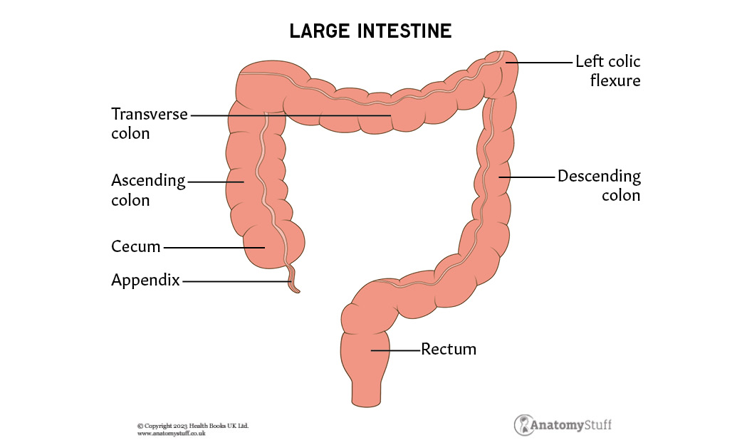
The large intestine begins at the ileocecal junction, a sphincter muscle situated at the junction of the ileum and the colon. It contains several unique structures which help to differentiate it from the small intestine:
• Omental appendices – Small pouches of the peritoneum situated along the colon and upper part of the rectum
• Haustra – Small pouches which give the large intestine a segmented appearance
• Taeniae coli – Three bands of longitudinal smooth muscle on the colon surface
It is also shorter and wider than the small intestine.
Accessory organs
The salivary glands, liver, gallbladder, and pancreas are accessory organs. This is because they are not part of the digestive tract but aid the processes of ingestion, digestion, and absorption.
| Accessory organ | Function |
| Salivary glands | They are three main salivary glands: parotid, submandibular, and sublingual glands (all paired). Saliva contains water, mucous and the enzyme amylase. It has many important roles, such as cleansing the teeth, lubricating food during mastication and swallowing and initiating the chemical digestion of starches through the action of amylase. |
| Liver | The liver has many important roles, such as the synthesis of bile salts (which help with the digestion and absorption of fat) and carbohydrate, lipid and protein metabolism. |
| Gallbladder | The gallbladder stores a reservoir for bile, which is primarily made up of water, bile salts, bile pigments, and cholesterol. |
| Pancreas | Pancreatic enzymes help digestion by breaking down fats, proteins and carbohydrates. |
Blood supply
The major arteries supplying the gastrointestinal tract are the celiac, superior mesenteric, and inferior mesenteric arteries.
The celiac supplies the stomach and the duodenum. The superior mesenteric supplies the rest of the small intestine and the proximal portion of the colon, while the inferior mesenteric supplies the distal portion of the colon.
Innervation
The GI tract is innervated by the sympathetic nervous system, parasympathetic nervous system and enteric nervous system.
The enteric nervous system is unique to the GI tract and regulates digestive processes within the gastrointestinal tract. It is made up of over one hundred million neurones found within the lower oesophagus all the way to the rectum. It also includes two plexuses (myenteric and submucosal) and their associated ganglia.
Embryological origin
Each organ in the GI tract develops from one of either the foregut, midgut or hindgut:
• The foregut gives rise to the trachea, lungs, oesophagus, stomach, liver, gallbladder, pancreas and upper duodenum
• The midgut gives rise to the lower duodenum, jejunum, ileum, cecum, appendix, ascending colon and proximal 2/3 of the transverse colon.
• The hindgut gives rise to the distal 1/3 of the transverse colon, descending colon, sigmoid colon and rectum.
In conclusion, the gastrointestinal tract is a remarkable and intricate system that plays a pivotal role in our overall health and well-being. We have learnt all the organs involved which to recap is the oral cavity, pharynx, oesophagus, stomach, small intestine and large intestine.

Related Products
View All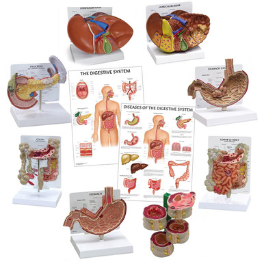

AnatomyStuff
The Anatomy of Irritable Bowel Syndrome (IBS) Chart / Poster - Laminated
Now
£14.00
Inc VAT

AnatomyStuff
Pharynx & Larynx Anatomy Revision Worksheets (Interactive & Printable PDFs)
Now
£5.00
Inc VAT


