Introduction
The integumentary system serves as the anatomic boundary between the body and the external environment. It consists of the skin, hair, nails, and sebaceous glands that produce sweat and oil. Keep reading to learn all about its anatomy, including its blood supply, innervation and embryological origin. Learn even more by checking out our Hair & Skin Anatomy Revision Worksheet.
Function of the skin
The skin, which is considered to be the largest organ in the human body, is an integral part of the integumentary system. It acts as a barrier against external substances such as water, microorganisms, toxins and UV light. It also has many other important roles like body temperature regulation, cell fluid maintenance, synthesis of Vitamin D and detection of stimuli. Additionally, its general appearance can provide an important insight into the patient’s overall health, such as their hydration status.
Layers of the skin
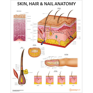
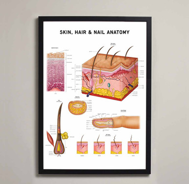
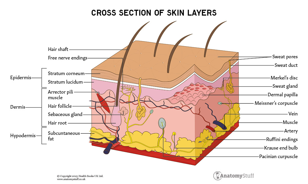
The skin consists of three main layers, which all vary significantly in their anatomy and function:
• Epidermis
• Dermis
• Hypodermis (also known as subcutis or subcutaneous fat)
Epidermis
The epidermis is the outermost layer of the skin. It acts as a waterproof barrier and contributes to the skin tone. It contains different various cell types:
| Cell type | Function |
| Keratinocytes
|
Produce keratin, a fibrous protein
Regulate calcium absorption |
|
Melanocytes
|
Produce melanin |
|
Langerhans’ cells
|
Part of the immune system |
|
Merkel’s cell |
Touch receptors |
Keratinocytes are the most abundant cell type in the epidermis. The protein they produce, keratin, helps form the tissues of the hair and nails.
Melanin is a substance present in the skin that produces pigment. Each person has varying amounts of melanin in their skin.
The epidermis also has several different layers (from deep to superficial):
• Stratum basale (also known as the basal cell layer or the stratum germinativum)
• Stratum spinosum (also known as the squamous cell layer)
• Stratum granulosum
• Stratum lucidum
• Stratum corneum
The stratum basale, or basal layer, contains melanocytes and small round cells called basal cells. The basal cells continuously divide, and the new cells push the old ones to the skin’s surface.
The stratrum spinosum is also known as the squamous cell layer. It contains basal cells that have been pushed upward from the basal layer. These are now called keratinocytes. This thick layer also contains the Langerhans’ cells which alert the immune system about the presence of antigens.
The keratinocytes are then pushed up the stratum granulosum and the stratum lucidum layers. They eventually become dehydrated and die.
The stratum corneum is the outermost layer of the epidermis. It contains many thin layers of continually shedding, dead keratinocytes. As the outermost cells age and wear down, they are replaced by new layers of strong cells. This shedding process slows down with time and takes approximately 40 days.
All five layers of the epidermis can be found on areas of thick, hairless skin like the palms of the hands and soles of the feet. In other places, the epidermis lacks the stratum lucidum and, therefore, only has four layers.
The basal layer is separated from the dermis by a highly specialised structure called the basement membrane, which is also known as the basal lamina.
Dermis
The dermis, which is located beneath the epidermis, is the thickest layer of skin. As mentioned above, it is connected to the epidermis at the level of the basement membrane. It can be divided into two layers (from superficial to deep):
• Papillary dermis
• Reticular dermis
The papillary layer comprises thin layers of collagen fibres, whereas the reticular dermis consists of thick collagen fibres.
In terms of their function, the papillary layer supplies nutrients to select layers of the epidermis and regulates temperature. The reticular layer provides structure and elasticity to the skin.
Hypodermis
The hypodermis, also known as the subcutis or subcutaneous layer, is the innermost layer of the skin. It lies directly below the dermis and consists of a network of fat and collagen cells. The stored fat is present in cells called adipocytes and has several important functions, including:
• Energy reserve
• Insulator
• Shock absorber
Other important structures like blood vessels, nerves, lymph vessels, and hair follicles also cross through this layer.
Specialised cells and structures
The skin contains various specialised structures:
| Specialised cells and structures | Function |
| Nails | The nail bed is a specialised structure of the epidermis that is found at the tips of the fingers and toes
|
| Hair follicles | A tube-shaped sheath surrounds and nourishes the hair
|
| Sebaceous glands | Located alongside hair follicles and is found everywhere apart from the palms of the hands and the soles of the feet. They secrete an oily, waxy substance called sebum which forms a protective coating on the skin.
|
| Sweat glands | There are two types: apocrine glands and eccrine glands.
Apocrine glands are specialised sweat glands found in the armpits, groin, pubic region and area around the breast. They encourage the growth of the bacteria responsible for body odour. Eccrine glands are found all over the body, and they secrete mostly water.
|
Each hair follicle is attached to a tiny muscle called the arrector pili muscle. This band of smooth muscle connects the hair follicle to the connective tissue of the basement membrane. When they contract, the hairs stand up, and this is known as goosebumps.
The hair follicle, arrector pili muscle and sebaceous gland are collectively known as the pilosebaceous unit. All of the components of the pilosebaceous unit are under endocrine control.
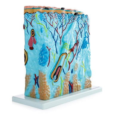
Blood supply
The skin is highly vascularised and is supplied by plexuses found between the reticular and papillary layers of the dermis. The epidermis does not contain any blood vessels.
Innervation
The skin is a very sensitive organ. It contains touch receptors, which are a type of sensory neuron that allow us to perceive the changes in the external environment.
The touch receptors possess specialised endings that respond to mechanical stimulation and transmit this information to the central nervous system:
- Thermoreceptors detect temperature changes
- Nociceptors detect pain
- Mechanoreceptors detect physical change. These include:
- Pacinian corpuscles (detect deep pressure and vibrational changes)
- Meissner corpuscles (detect light touch)
- Ruffini corpuscles (detect skin stretching and finger position)
- Merkel complexes (gentle touch sensation)
- C-fibre LTM (low threshold mechanoreceptors)
Embryology
The epidermis is derived from ectodermal tissue, and the dermis and hypodermis are derived from mesodermal tissue.
Refresher
A germ layer is a group of cells in the embryo that differentiate into different tissues and organs. The three germ layers are the endoderm, the ectoderm and the mesoderm.
In wrapping up our comprehensive revision guide on hair & skin anatomy, we’ve delved into the intricate structures of these two essential components of our bodies. We’ve explored how sensitive our body’s largest organ the skin is and how good it is at shielding us from the outside world and regulating numerous bodily functions.