


![]()
Introduction
The hand is the most distal part of the upper limb and is connected to the body at the wrist joint. The front of the hand is referred to as the palmar side, and the back as the dorsal side. The anatomy of the hand and wrist is intricate, delicate and unique. It is composed of an incredible arrangement of bones, muscles and ligaments which work together and enable us to grip, grasp and form precise movements. Unlike other primates, humans have longer thumbs and flexible fingers, and this allows us to perform a wide range of complex actions with flexibility and dexterity.
Joint Type
The Wrist Joint
The wrist is the site at which the forearm connects to the arm. In anatomical terms, the wrist joint is referred to as the radiocarpal joint. This is because the distal radius of the forearm and articular disc articulates with the proximal row of carpal bones in the hand (except pisiform).
Whilst the forearm consists of both ulnar and radial bones, it is important to note that only the radius is involved in the formation of the wrist joint. Close to the wrist joint, the ulna articulates with the radius beside it, but a structure known as the triangular fibrocartilaginous complex prevents the ulna from directly connecting to the carpal bones. This complex lies over the superior ulna surface and allows for load-bearing at the wrist.
Structurally the radiocarpal joint is classified as a synovial joint which means that it is enclosed by a dual-layered capsule. The inner membrane of the joint capsule secretes synovial fluid, and this allows for the lubrication of the joint. Functionally, the radiocarpal joint is classified as an ellipsoidal joint, meaning that movement occurs biaxially i.e., along two axes. The movements at the wrist joint are flexion, extension, adduction, and abduction.
The Hand Joint
The hand itself consist of several smaller joints known as interphalangeal joints. These joints provide the hand with fine motor function. They are all classified as synovial hinge joints meaning that movement occurs in one plane, like the hinge of the door. These movements are flexion and extension.
The fingers have three joints, and the thumb has two joints, and these are:
• metacarpophalangeal joint (MCP) – the joint at the finger base i.e., the knuckle
• proximal interphalangeal joint (PIP) – the joint in the middle of the finger
• distal interphalangeal joint (DIP) – the joint closest to the fingertip.
Interestingly, the strength of the movement performed by the finger joints is dependent on the position of the wrist joint. When the wrist is extended, the flexors of the fingers have the most powerful leading to a strong grip. Wrist flexion produces a weak grip.
Osteology
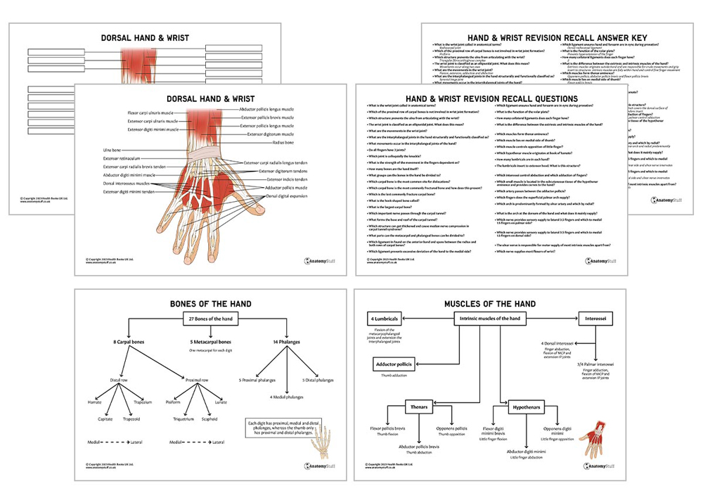
With a total of 27 bones in such a small area, the hand has an incredibly complex skeleton. The bones of the hand can be divided into 8 carpal bones (some of which form the wrist joint), 5 metacarpal bones and 14 phalangeal bones. The carpal bones of the hand meet the radius (and ulna indirectly), forming the wrist joint.
8 Carpal Bones
The carpal bones are divided into two rows: proximal and distal.
From medial to lateral (pinkie to thumb), the proximal row consists of the:
pisiform
• small round pea-shaped bone in the flexor carpi ulnaris tendon
• not involved in movements of the wrist
• easily palpable at the medial base of the palm as lies superficially
triquetrum
• pyramid-shaped
• fractures of the bone occur in forced dorsiflexion
lunate
• crescent/ moon shaped
• the most common site for dislocations compared to other carpal bones
scaphoid
• the largest carpal bone of the proximal row
• most commonly fractured carpal bone- presents as pain and tenderness in the anatomical snuff box region
Apart from the pisiform, the rest of the carpal bones in the proximal row are involved in the formation of the radiocarpal joint.
From medial to lateral the distal row consists of the:
hamate
• hook-shaped bone
• articulates with the 5th metacarpal e.g., little finger
capitate
• largest carpal bone
• articulates with the 3rd and 4th metacarpal e.g., middle and ring finger
trapezoid
• four-sided and similar in shape to the trapezium
• least commonly fractured carpal bone
• articulates with the 2nd metacarpal e.g., index finger
trapezium
• four-sided and similar in shape to the trapezius finger
• articulates with the 1st metacarpal e.g., thumb
Carpal Tunnel
The carpal bones are arranged in a U-shape and form the carpal tunnel. This is a narrow passageway for several tendons and the median nerve to enter the wrist. Located at the anterior wrist, the base of the carpal tunnel is formed by a deep concave carpal arch and the roof by superficial flexor retinaculum. These structures form a bridge between scaphoid and trapezium tubercles laterally and pisiform and hook of hamate medially. The flexor retinaculum is a thick connective tissue which can get thickened and cause median nerve compression in carpal tunnel syndrome.
5 Metacarpal Bones
These bones form the most proximal part of the thumb and fingers. They articulate with the distal row of the carpal bones proximally and phalanges distally to form the metacarpophalangeal joint. The metacarpal bones can be divided into a head, body and base. All the metacarpals also articulate with each other apart from the thumb, and this allows the thumb to move freely and independently.
14 Phalanges
Like the metacarpal bones, each phalange can be divided into a head, body and base. The thumb has proximal and distal phalanges, whereas the other fingers have a proximal, middle and distal phalange. The phalanges articulate to form the proximal and distal interphalangeal joints.
![]()
Ligaments
The complex arrangement of the joints in the hand and wrist are supported by numerous ligaments. The main ligaments supporting the fingers are the collateral ligaments and the volar plate.
The Wrist Ligaments
The 4 main ligaments of the wrist joint are:
Palmar radiocarpal ligament – found on the anterior hand and spans between the radius and both rows of carpal bones. It provides stability and ensures the forearm and hand are in sync during supination.
Dorsal radiocarpal ligament – found on the posterior hand and spans between the radius and both rows of carpal bones. It provides stability and ensures the forearm and hand are in sync during pronation.
Ulnar collateral ligament – runs from the styloid process of the ulna to the triquetrum and pisiform. It prevents excessive deviation of the hand to the lateral side.
Radial collateral ligament – runs from the styloid process of radius to the scaphoid and trapezium. It prevents excessive deviation of the hand to the medial side.
The Hand ligaments
The main ligaments supporting the small joints in the fingers are:
Collateral ligaments – these are two ligaments on either side of each finger that prevents abnormal sideway hyperextension. They span from the head of the metacarpal bone to the proximal phalanx.
Volar plate – A thickened ligament at the palmar metacarpophalangeal joint which prevents hyperextension of the finger.
Musculature and Movement
Not only do the hand and wrist allows us to powerfully grip and grasp objects, but we are also able to perform precise and delicate movements with extreme control.
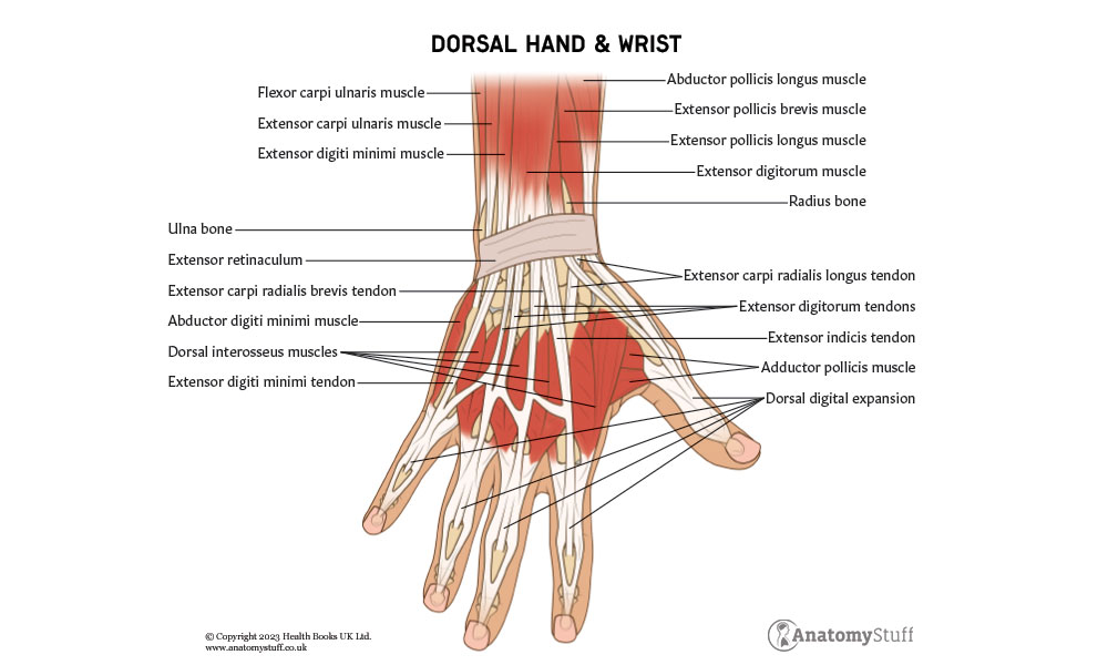
The hand and wrist muscles can be divided into extrinsic and intrinsic muscles. The extrinsic muscles originate outside the hand, most being in the forearm, and insert into structures of the hand. They are responsible for strong grip and crude movements. The intrinsic muscles are located within the hand and carry out fine motor functions.
Muscles acting on the Wrist Joint
All movements occurring in the wrist joint are produced by the muscles in the anterior and posterior compartments of the forearm:
Flexion
• flexor carpi ulnaris
• flexor carpi radialis
• flexor digitorum superficialis
Extension
• extensor carpi radialis longus and brevis
• extensor carpi ulnaris
• extensor digitorum
Adduction
• extensor carpi ulnaris
• flexor carpi ulnaris
Abduction
• abductor pollicis longus
• flexor carpi radialis
• extensor carpi radialis longus and brevis
Intrinsic Muscles of the Hand
The intrinsic muscles of the hand can be divided into thenar, hypothenar, lumbricals, interossei and others.
Thenar Muscles
There are 3 thenar muscles at the base of the thumb, and these are responsible for thumb movement. Their muscle bellies form the thenar eminence.
Opponens pollicis – largest and lies deep to the other two
• Origin at tubercle of trapezium
• Insertion to the lateral side of the first metacarpal
• Controls thumb opposition by medial rotation and flexion of the first metacarpal
Abductor pollicis brevis – superficial and on the lateral side
• Origin at tubercle of scaphoid and trapezium
• Insertion at the lateral side of thumb proximal phalanx
• Controls abduction of the thumb
Flexor pollicis brevis – superficial and on medial side
• Origin at tubercle of trapezium
• Insertion at the base of thumb proximal phalanx
• Controls flexion of the thumb
Hypothenar Muscles
There are 3 hypothenar muscles at the base of the pinkie, and these are responsible for little finger movement. Their muscle bellies form the hypothenar eminence.
Opponens digiti minimi – lies deep to the other two muscles
• Origin at the hook of hamate
• Insertion at the medial side of the 5th metacarpal
• Controls opposition of little finger by rotation towards the palm
Abductor digiti minimi –superficial on the medial side
• Origin at pisiform
• Insertion at the lateral side of little finger proximal phalanx
• Controls abduction of the little finger
Flexor digiti minimi brevis – superficial and on the lateral side
• Origin at the hook of hamate
• Insertion at the base of little finger proximal phalanx
• Controls flexion of the little finger
Lumbricals
There are 4 lumbricals in each hand, and these are responsible for fine finger control. Each lumbrical span from the tendon of the flexor digitorum fundus travels dorsally and laterally to each finger and inserts into the extensor hood. The extensor hood is a special connective tissue which covers the dorsal surface of the metacarpophalangeal joint where extensor tendons insert. The lumbricals control the flexion and extension of the metacarpophalangeal joint.
Interossei
The interossei muscles are located between the fingers. They can be subdivided into 4 dorsal and 3/4 palmar interossei. On their respective dorsal or palmar sides, the interossei originate from the lateral and medial surface of the metacarpals and insert into the proximal phalanx and extensor hood of each finger. The dorsal interossei control abduction and palmar control adduction. Both also assist in the flexion of the metacarpophalangeal joint and the extension of interphalangeal joints.
Other Muscles
The palmaris Brevis is a tiny thin muscle in the subcutaneous tissue of the hypothenar eminence. It provides curves to the hand at this location, supporting the grip function.
The adductor pollicis is a large bicipital muscle which is triangular in shape. Its heads originate from metacarpals and insert to the base of the thumb of the proximal phalanx. This muscle forms the deep palmar arch and controls the adduction of the thumb. The radial artery passes between two heads.
Vascular Supply
Arterial Supply
The wrist and hand are supplied by the radial and ulnar arteries and their branches which are arranged in anastomotic arch structures.
In the palm of the hand, the radial and ulnar arteries merge to form the superficial and deep palmar arches.
• The superficial palmar arch is predominantly formed by the ulnar artery and has a small contribution from the radial artery. It provides blood supply to the ulnar half of the index finger, 3rd, 4th and 5th fingers via the common digital artery and its branches.
• The thumb and radial half of the index finger are supplied directly by the radial artery before it goes on to form the deep palmar arch.
• The deep palmar arch is predominantly formed by the radial artery with a small contribution form the ulnar artery. It supplies the deep structures of the hand.
In the dorsum of the hand, the radial and ulnar arteries merge to form the dorsal carpal arch. The posterior and anterior interosseous arteries also contribute. The branches of these arch run across metacarpal bones and supply the wrist.
Venous Drainage
The venous drainage of the hand and wrist is carried out by paired veins which run alongside the arterial arches. The dorsum of the hand is drained by the superficial cephalic and basilic veins.
Nerve Supply
The main nerves innervating the hand and wrist are the median, ulnar and radial nerves. They provide both the sensory and motor supply in different distributions.
Radial Nerve
• sensory supply of the lateral 2/3 of the dorsum of the hand and the dorsal surface of the lateral 3.5 fingers sensory branch.
• motor supply to most of the extensor muscles in the forearm.
Ulnar Nerve
• sensory supply to both the palmar and dorsal side of the medial 1.5 fingers.
• motor supply to the ulnar half of the flexor digitorum profundus, flexor carpi ulnaris, as well as most of the intrinsic muscles of the hand (median nerve responsible for a few intrinsic muscles).
Median Nerve
• sensory supply to the palmar surface of the lateral 3.5 fingers as well as their nail beds dorsally via the proper digital nerves. It also supplies sensation to the lateral 2/3 of the palm of the hand via the palmar cutaneous branch.
• motor supply to most of the flexors of the wrist and hand. It also innervates the thenar eminence and the lateral two lumbricals.

Related Products
View All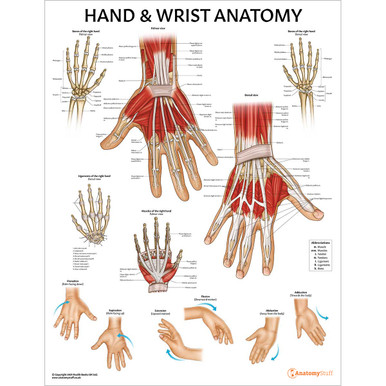

save
30%
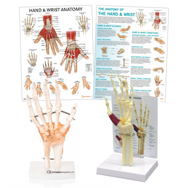
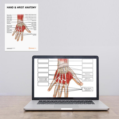
AnatomyStuff
Hand & Wrist Anatomy Poster / Worksheet (Interactive & Printable PDF)
Now
£3.50
Inc VAT
Was
£5.00
Inc VAT


