


![]()
Introduction
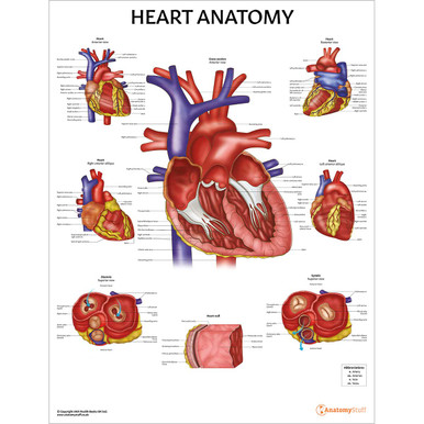
The heart is an important organ of the cardiovascular system. It pumps blood to the entire body to ensure proper oxygen and nutrients are delivered to cells. The human heart is roughly the size of a fist. The heart is located in the middle mediastinum, a section of the thorax that is located between the two lungs and is anterior to the vertebrae.
Walls of the Heart
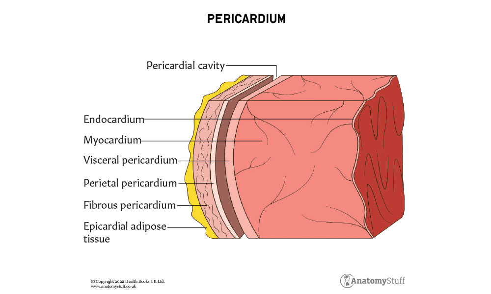
Each heart chamber is surrounded by walls that consist of three distinct layers. From deep to superficial, the layers are:
Endocardium – the innermost layer of connective tissue that lines the chambers and valves of the heart. This is continuous with the large blood vessels that enter the heart
Myocardium – thick, muscular middle layer that consists of cardiac myocytes arranged in overlapping spiral patterns
Epicardium – thin superficial layer that forms part of the visceral layer of the serous pericardium
Right side of the Heart
Poorly oxygenated blood from the body drains into the right atrium through two large vessels, namely superior vena cava (SVC) and inferior vena cava (IVC). This travels down the right side of the heart into the right ventricle and is pumped to the lungs through the pulmonary trunk and arteries.
Features of the right side of the heart:
Right atrium:
• Right auricle – cone-shaped pouch attached to the right atrium
• Corresponds to the right pulmonary surface of the heart
• Anterior wall is composed of pectinate muscles (rough muscular ridges)
• Crista terminalis – thickened band from which the pectinate muscles arise present in the walls of the right atrium
• Opening of the coronary sinus – drains venous blood from the cardiac muscle into the right atrium
• Oval fossa – a remnant of the oval foramen (present in the foetus). This is an opening between the right and left atria.
Right ventricle:
• Corresponds to the anterior (sternocostal) surface
• Conus arteriosus (infundibulum) – a cone-shaped structure from which the pulmonary trunk and arteries emerge
• Trabeculae carneae – irregular muscular elevations present on the inner walls of the right and left ventricles
• Tricuspid valve – prevents the backflow of blood travelling from the right atrium to the right ventricle
• Tendinous chords – attach to the cusps of the tricuspid valve and prevent prolapse of the valve
• Papillary muscles – muscle projections from the right ventricular walls that give rise to the tendinous chords
Left side of the Heart
Well oxygenated blood from the lungs enters the left atrium through the pulmonary veins. This blood travels to the left ventricle, where with the help of the muscular wall of the left ventricle, blood is pumped through the aorta for distribution to the entire body.
Left atrium:
• Left auricle – pouch attached to the left atrium and lined with pectinate muscles
• Openings to four pulmonary veins – two superior pulmonary veins and two inferior pulmonary veins
Left ventricle:
• Corresponds to the left pulmonary surface and forms the cardiac impression of the left lung
• Walls of the left ventricle are nearly three times thicker than the right ventricle
• Trabeculae carneae – irregular muscular elevations present on the inner walls of the right and left ventricles. They are present in greater quantity in the left ventricle
• Mitral valve – prevents the backflow of blood travelling from the left atrium to the left ventricle
• Tendinous chords – attach to the cusps of the mitral valve and prevent prolapse of the valve
• Papillary muscles – muscle projections from the left ventricular walls that give rise to the tendinous chords
![]()
Valves of the Heart
There are 4 main valves in the heart. The function of a valve is to prevent the backflow of blood from one chamber to another. Cusps are folds of a valve that allow the valve to be open or closed. They are extended from tendinous cords and are controlled with the help of papillary muscle attachments.
Tricuspid valve:
• Between the right atrium and ventricle
• Consists of 3 cusps – septal, anterior and posterior
Pulmonary valve (semilunar)
• Between the right ventricle and pulmonary artery
• Known as a semilunar valve because it is shaped like a half moon
• Consists of 3 cusps – anterior, right and left
Mitral valve (bicuspid)
• Between the left atrium and ventricle
• Consists of 2 cusps – anterior and posterior
Aortic valve (semilunar)
• Between the left ventricle and aorta
• Known as a semilunar valve because it is shaped like a half moon
• Consists of 3 cusps – posterior, right and left
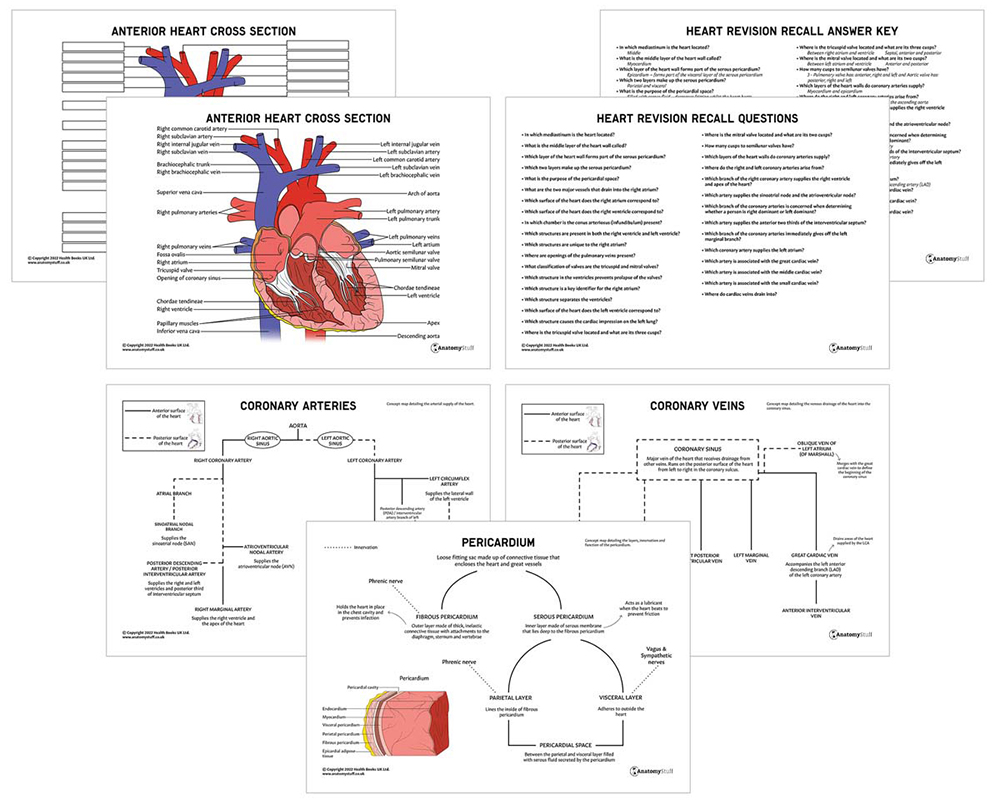
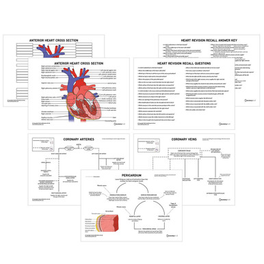
Arterial supply of the Heart
Coronary arteries are branches of the aorta and supply the myocardium and epicardium. Right coronary artery (RCA) and left coronary artery (LCA) emerge from the aortic sinus and branch off to give many further arteries to supply the heart muscle.
Right coronary artery
• Arises from the right aortic sinus of the ascending aorta
• Branches of RCA:
• Sinoatrial nodal artery – supplies the sinoatrial node (SAN)
• Right marginal artery – supplies the right ventricle and apex of the heart
• Posterior descending/ interventricular artery – supplies the right and left ventricles and the posterior third of the interventricular septum (IVS)
• Atrioventricular nodal artery (arises from near the origin of the posterior descending artery in the RCA) – supplies the atrioventricular node (AVN)
Left coronary artery
• Arises from the left aortic sinus of the ascending aorta
• Branches of LCA:
• Left anterior descending/ anterior interventricular artery – supplies the right and left ventricles and anterior two-thirds of the interventricular septum (IVS)
• Left diagonal branches are given off by the LAD – supplies the left atrium and anterior left ventricle
• Left circumflex artery – supplies the lateral wall of the left ventricle
• Left marginal branch is given off by the left circumflex artery – supplies the left ventricle
Venous drainage of the Heart
All veins of the heart drain into the coronary sinus, a larger vein that runs along the posterior surface of the heart and drains into the right atrium.
The coronary sinus receives drainage from:
• Great cardiac vein (anterior interventricular) – originates from the apex of the heart and travels superiorly to join the coronary sinus. This vein is associated with the left anterior descending artery (LAD)
• Middle cardiac vein – originates from the apex and ascends in the posterior interventricular groove to join the coronary sinus. This vein is associated with the right posterior descending/ interventricular artery
• Small cardiac vein – located on the anterior surface of the heart and travels in a groove between the right atrium and ventricle to reach the posterior surface of the heart where it joins the coronary sinus. This vein is associated with the right marginal artery.

Related Products
View All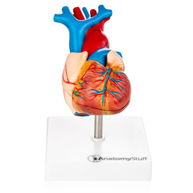
AnatomyStuff
Heart Anatomy Revision Worksheets (Interactive & Printable PDFs)
Now
£5.00
Inc VAT


