


![]()
The thoracic cavity which is surrounded by the thoracic wall has three main compartments: one central compartment called mediastinum (contains the heart and its vessels, trachea, oesophagus and thymus); and two lateral compartments that contain each lung and its pleura.
Pleura and Pleural Cavities
There are two pleural cavities that surround the lungs. The space between the visceral pleura and parietal pleura is known as the pleural cavity and contains a thin layer of serous fluid. Each pleura is composed of single layer flat cells known as mesothelium and an associated layer of connective tissue.
Each lung is lined by two layers of serous membrane known as pleurae. These layers are the outer parietal pleura and the inner visceral pleura. The space between the pleura is known as the pleural cavity and contains a thin layer of serous fluid. Each pleura is composed of single layer flat cells known as mesothelium and an associated layer of connective tissue.
• Visceral pleura – this structure is directly attached and covers the lungs
• Parietal pleura – this structure is attached to the thoracic wall, mediastinum and diaphragm. Parietal pleura can be further divided based on its anatomical location:
◦ Costal part – covers the surfaces of the thoracic wall (ribs, sternum, costal cartilages etc.)
◦ Diaphragmatic part – covers the superior surface of the diaphragm
◦ Mediastinal part – covers the lateral surface of the mediastinum
Cervical part/pleura cupola – extends through the superior thoracic aperture into the root of the neck. It forms a dome over the apex of the lungs.
Suprapleural membrane is a connective tissue membrane that covers the cervical pleura. This membrane provides apical support to the pleural cavity in the root of the neck.
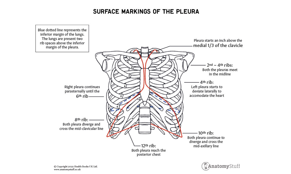
Recesses
During expiration, lungs deflate and do not completely occupy the pleural cavities. This leads to the formation of areas where the two adjacent layers of parietal pleura come in contact and are only separated by serous fluid.
There are two main pleural recesses and these include:
• Costomediastinal recess
◦ This is smaller of the recesses and lies behind the sternum
◦ At this location, the costal pleura is in contact with the mediastinal pleura
◦ The left costomediastinal recess is less occupied than the right due to the presence of the cardiac notch on the left lung
• Costodiaphragmatic recess
◦ At this location, the costal pleura is in contact with the diaphragmatic pleura
◦ These recesses are deepest after forced expiration and shallowest after forced inspiration
Features of the Lungs
There are two lungs that lie either side of the mediastinum. They act as vital organs of respiration by oxygenating the blood and removing carbon dioxide from it. This process occurs through gas exchange. Oxygenated blood from the lungs enters the heart via pulmonary veins and deoxygenated blood from the heart is transported to the lungs via pulmonary arteries. Each lung has the following features:
• Apex
◦ This is the features that projects above the first rib and into the root of the neck. It is the superior-most feature of the lungs
◦ Covered by cervical pleura
◦ Base/diaphragmatic surface
◦ This is the inferior surface of the lungs that sits on the diaphragm
• Surfaces
◦ Costal surface – immediately posterior to the ribs and intercostal spaces
◦ Mediastinal surface – contains the hilum and lies anteriorly against the mediastinum
• Borders
◦ Inferior border – separates the diaphragmatic surface from the costal and mediastinal surfaces
◦ Anterior and posterior border – separates the costal surface and medial surface
◦ The anterior and inferior borders are sharp whereas the posterior border is smooth
Right and Left Lung
Each lung contains identifiable lobes and fissures. The right lung has two fissures that separate the lung to make three lobes. These lobes are have free movement against each other as they are separated. The visceral pleura invaginates along the fissures. Each lung also contains a root which is made up of vessels that enter and leave the lungs. These structures are covered by a reflection of the pleura called the hilum.
• Fissures
◦ The two fissures of the right lung are the oblique fissure and the horizontal fissure and these separate the right lung into three lobes:
■ Oblique fissure separates the inferior and superior lobe
■ Horizontal fissure separates the superior lobe from the middle lobe
◦ The left lung is separated into the superior and inferior lobe by the oblique fissure
• Root and hilum structures
◦ Bronchus and Pulmonary artery
■ In the right lung, the bronchus is superior to the pulmonary artery
■ In the left lung, the bronchus is inferior to the pulmonary artery
◦ Bronchial vessels
◦ Two pulmonary veins
◦ Nerves
◦ Lymphatics
• Pulmonary ligament
◦ This is a projection of the pleura on both the right and left lung that runs inferiorly to form the root of the lung. It helps to stabilise the inferior lobe.
![]()
Bronchial Tree
The lower respiratory system is made up of cartilaginous tube called the trachea that extends from vertebral level C6 to T5. It divides into many branches as it extends inferiorly into the thorax. The bronchial tree distributes air to the lungs and allows proper ventilation of the body.
• Trachea
◦ A C-shaped transverse hyaline cartilage that helps to keep the trachea open and prevent collapse during expiration
◦ The lumen of the trachea is kept open by around 20 tracheal cartilaginous rings. The gaps between these cartilaginous rings is filled by the trachealis muscle (smooth muscle).
■ This allows flexibility of the trachea during inspiration and expiration
• Bronchi
◦ The trachea bifurcates and gives rise to two main primary bronchi which divide further and branch into secondary (lobar) bronchi and then tertiary (segmental) bronchi.
◦ Going down the bronchial tree the amount of cartilage decreases and the amount of smooth muscle increase
◦ The tertiary bronchi contains the least cartilage and the most smooth muscle
• Bronchioles
◦ Tertiary bronchi give rise to the bronchioles.
◦ The bronchioles have a cartilage-free wall and are surrounded by smooth muscle
◦ The bronchioles can be divided by size into the larger lobular bronchioles, smaller terminal bronchioles and the smallest respiratory bronchioles.
◦ The mucus membrane lining of the lobular bronchioles is made up of ciliated columnar epithelium
◦ At the respiratory bronchioles the mucus membrane lining changes to simple cuboidal epithelium
◦ The respiratory bronchioles lead to alveoli. These are tiny air sacs where gas exchange takes place and this is the final part of the bronchial tree.

Related Products
View All

save
30%
AnatomyStuff
Lung Anatomy Poster / Worksheet (Interactive & Printable PDF)
Now
£3.50
Inc VAT
Was
£5.00
Inc VAT

AnatomyStuff
Lung Anatomy Revision Worksheets (Interactive & Printable PDFs)
Now
£5.00
Inc VAT


