


![]()
Introduction
The lymphatic system is a network of vessels, nodes, and ducts that collect and circulate excess fluid in the body. While often underappreciated, it is an integral component of the circulatory, immune, and metabolic systems. Its main functions include the enhancement of the immune system, absorption of dietary fats and fluid balance maintenance. This revision guide will explore the anatomy of this complex system and its important role in the human body.
Lymph
Lymph is a pale fluid that flows throughout the lymphatic system. It originates from the fluid that has leaked from the vascular system into the interstitial space. It is then reabsorbed via the lymphatic capillaries. Interestingly, approximately 10% of blood volume becomes lymph, and a healthy adult will produce around 3-4 litres of lymphatic fluid every single day.
Lymphatic Vessels
The lymphatic transport system can be subdivided into many different components: capillaries, collecting vessels, afferent lymph cells, lymph nodes, efferent lymph vessels and ducts:
1. The lymph capillaries are thin-walled, microscopic vessels found in almost all regions of the body. Because they are single-ended, fluid is able to enter the capillary but not able to leave.
2. The lymph capillaries drain into larger collecting lymphatics. These vessels transport lymph using smooth muscle walls (which contract to propel the lymph forward) and smooth muscle walls (which contract to propel the lymph forward), and valves to prevent the lymph from flowing backwards.
3. As the collecting lymph vessel accumulates more and more lymph, it eventually becomes an afferent lymph vessel as it enters a lymph node.
4. The lymph exits the lymph node via an efferent lymph vessel.
5. An efferent lymph vessel may directly drain into either the right or thoracic lymph ducts or into another lymph node. The right lymphatic duct drains lymph from the upper right quadrant of the body. The thoracic duct drains the rest.
6. The right lymphatic. The thoracic duct return lymph to the bloodstream by draining into the right and left subclavian veins.
Interesting fact!
For a long time, it was thought that the central nervous system (brain and spinal cord) was one of the few regions within the body did contain no lymphatic vessels. However, in 2015, a genuine and functional lymphatic vascular system was discovered in the meninges (the dura mater, the arachnoid mater, and the pia mater). More research is needed to fully understand the anatomy of these newly discovered lymphatic vessels.
![]()
Lymphatic vs Circulatory System
It is important to be aware of the key differences between the lymphatic and circulatory systems:
| Lymphatic system |
Circulatory system |
| · Defends against foreign microorganisms
· Absorption of dietary fats · Fluid balance (carries interstitial fluid back to the systemic circulation) |
· Delivers oxygen, nutrients and hormones to cells
· Removes waste products |
| Carries lymph | Carries blood |
| Lymphatic capillaries are larger and have closed ends (only carry fluid away from the tissues) | Blood capillaries are smaller and open-ended (open at one end to the arterioles and the other end to the venules) |
| Lymphatic diseases include lymphedema and lymphoma | Cardiovascular diseases include heart failure and myocardial infarction |
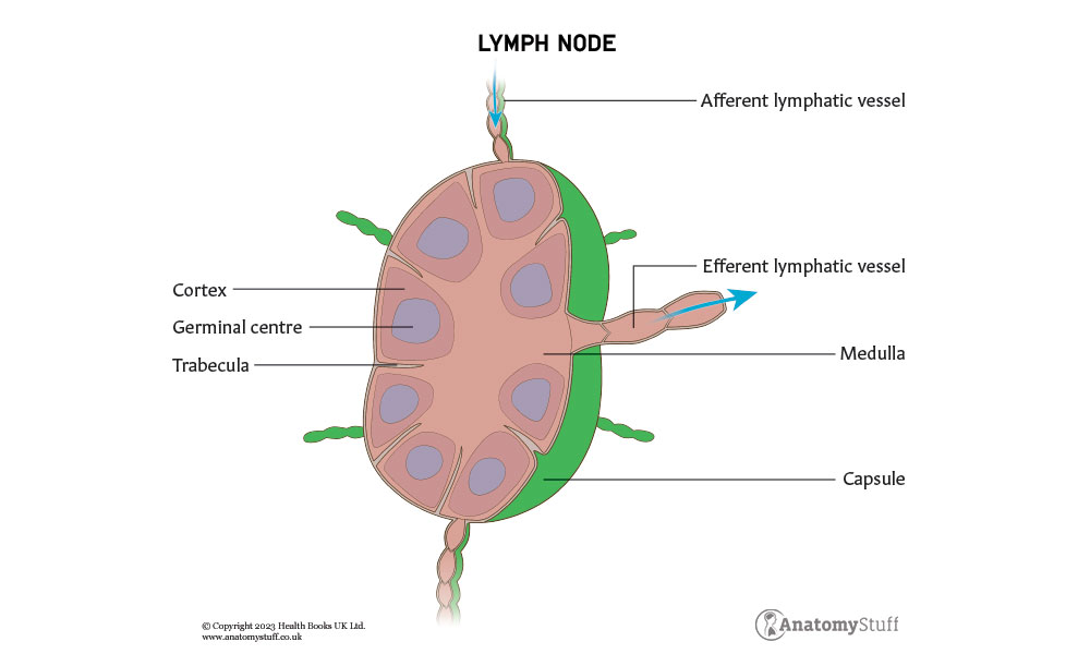
Lymph Nodes
A lymph node is a kidney-shaped organ, which is about 2cm in size, found at the convergence of major blood vessels. Every adult has approximately 800 lymph nodes which are mainly situated in the neck, axilla, thorax, abdomen and groin:
The lymph nodes in the head and neck are paired and broadly split into superficial and deep nodes:
| Superficial lymph nodes of the head and neck | Where do they drain lymph from? |
| Occipital nodes |
Skin overlying the occiput
|
| Mastoid (post-auricular) nodes | Posterior neck, the superior portion of the external ear, and the ear canal until the tympanic membrane |
| Pre-auricular nodes | Superficial face and temporal region |
| Superficial parotid nodes | Nose, the nasal cavity, part of the external ear canal, and the lateral orbit |
| Submental nodes | Middle-lower lip, the floor of the mouth, and the tip of the tongue |
| Submandibular nodes | Submental and facial nodes, the cheeks, the upper lip, and the marginal areas of the lower lip |
| Facial nodes | Mucous membranes of the inside of the cheek, the nasal mucosa, the eyelids, and the conjunctiva. |
| Superficial cervical nodes (divided into anterior superficial cervical and posterior superficial cervical) | Neck |
| Deep lymph nodes of the head and neck | Where do they drain lymph from? |
| Deep parotid nodes | Nasal cavity and the nasopharynx |
| Deep cervical nodes | Superficial nodes and all of the head and neck |
In the lower limb, the lymph nodes can be split into:
Inguinal Nodes – these nodes are further subdivided at the level where the great saphenous vein becomes the deep femoral vein. They become sub-inguinal lymph nodes below and superficial inguinal nodes above. Sub-inguinal nodes include superficial sub-inguinal nodes, deep sub-inguinal nodes and superficial inguinal nodes.
Iliac Nodes – these can be subdivided into external iliac nodes and common iliac nodes. External iliac nodes include the lateral external iliac lymph nodes, intermediate external iliac lymph nodes and medial external iliac lymph nodes. Common iliac nodes include lateral common iliac lymph nodes, intermediate common iliac lymph nodes and medial common iliac lymph nodes.
The lymph nodes of the upper limb include cubital, axillary, infraclavicular and interpectoral lymph nodes. These drain into the axillary nodes.
Each node is enclosed in a dense capsule with an indentation on one of its surfaces (hilum). Multiple afferent vessels (around 4 or 5) enter the lymph node through this hilum. The lymph is then filtered and leaves via one or two efferent vessels, which also exit via the hilum.
Within the lymph node, interstitial fluid from the soft tissues is filtered. By filtering the fluid, immune responses to pathogenic antigens are initiated (T-cells and B-cells are exposed to various antigens). B cells are mainly found in the outer cortex of the lymph node, whereas T cells and dendritic cells are mainly found in the paracortex.
During bacterial, viral, or fungal infections, autoimmune disease and malignancy, the lymph nodes may become swollen, known as lymphadenopathy.
Interesting fact!
While performing an abdominal examination, it is important to palpate for ‘Virchow’s Node’ – a supraclavicular node located in the left supraclavicular fossa (superior to the clavicle). Because it receives lymph drainage from the abdominal cavity, palpation of an enlarged Virchow’s node can indicate the presence of gastric cancer that has spread through the lymph vessels.
Lymphoid Organs
The lymphatic system also includes tissues and organs that make, store and release lymphocytes (a type of white blood cell which can be divided into B lymphocytes, T lymphocytes and natural killer (NK) cells). These lymphoid organs are classified as primary or secondary based on their function and structure.
There are two primary lymphatic organs: the red bone marrow and the thymus gland, which is where B and T lymphocytes form, multiply, and mature. More specifically, B lymphocytes mature within the marrow, and immature T lymphocytes migrate to the thymus for further development (which is why they are called T cells!)
Interestingly, with age, the thymus atrophies and declines in function. It is replaced with fatty tissue.
Secondary lymphatic organs include:
• Mucosa-associated lymphoid tissue (MALT) – located along mucosal linings throughout the body, including the tonsils, Peyer patches within the small intestine and the appendix
• Lymph nodes – located all around the body (mentioned above)
• Spleen – located in the left upper abdomen below the diaphragm
These secondary lymphoid organs provide the necessary environment for cells involved in the adaptive response to proliferate and mature.
Refresher
The immune system is made up of two parts: the innate immune system and the adaptive immune system. The adaptive immune response, which is carried out by two types of lymphocytes (B and T cells), is more complex than the innate and is triggered by pathogen exposure. It also involves immunological memory to learn about the threat and enhance the immune response accordingly. On the other hand, the innate immune system is more general and includes physical barriers (like the skin) as well as important cellular components such as K cells, macrophages, granulocytes, eosinophils, and antigen-presenting cells (dendritic cells).
Function
There are 3 primary functions of the lymphatic system:
1. Enhancement and facilitation of the immune system – Pathogens can easily enter this network to facilitate rapid spread throughout the body. To prevent this, the lymphatic system possesses filter-like structures (lymph nodes, as discussed above).
2. Maintenance of fluid balance – The lymphatic system returns excess interstitial fluid to the blood. Overall, about 90% is returned, and the remaining 10% becomes part of the interstitial fluid that surrounds the tissue cells.
3. Absorption of dietary fats from the gastrointestinal tract to the bloodstream for metabolism or storage – The mucosa that lines the small intestine is covered with finger-like projections called villi. In the centre of these villi, special lymph capillaries (called lacteals) are present, along with blood capillaries. Whilst the blood capillaries absorb most nutrients, the fats and fat-soluble vitamins are absorbed by the lacteals. Interestingly, the lymph in the lacteals is known as chyle due to its milky appearance caused by the high-fat content.

Related Products
View All
save
28%
AnatomyStuff
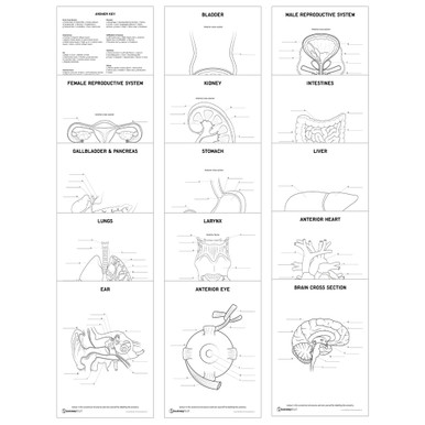
Major Organs of the Body 8 Chart Collection (Laminated)
Now
£80.00
Inc VAT
Was
£112.00
Inc VAT
AnatomyStuff
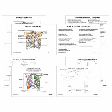
Thorax Anatomy Revision Worksheets (Interactive & Printable PDFs)
Now
£5.00
Inc VAT

AnatomyStuff
Lymphatic System Anatomy Revision Worksheets (Interactive & Printable PDFs)
Now
£5.00
Inc VAT


