


Introduction
The skull is a bony structure that encases the brain and supports the face. It is comprised of 22 individual bones and can be divided into two parts; the braincase (neurocranium) and the facial skeleton (viscerocranium). Keep reading to learn more about this important structure, including its bony landmarks and embryological origin.
To get the best revision experience, pair up this Skull Anatomy Revision Guide with our interactive Skull Anatomy Revision Worksheets.
Neurocranium
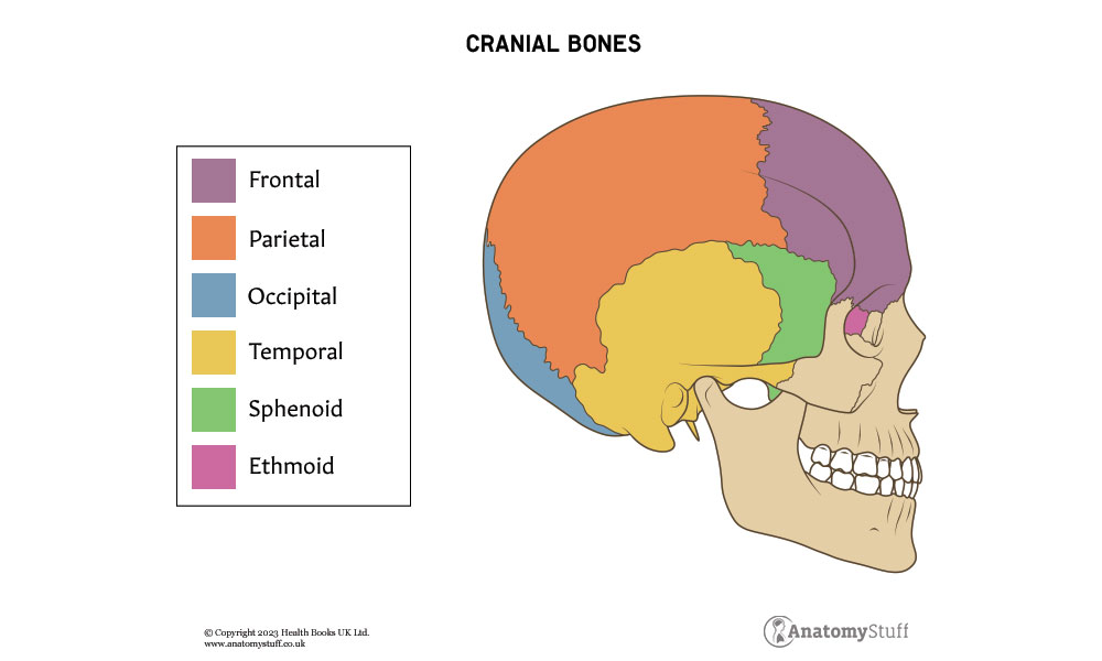
The neurocranium protects important structures such as the cerebral cortex, cerebellum, and orbital contents. Eight bones make up the neurocranium, including:
• Frontal bone
• Parietal bones (paired)
• Temporal bones (paired)
• Occipital bone
• Sphenoid bone
• Ethmoid bone
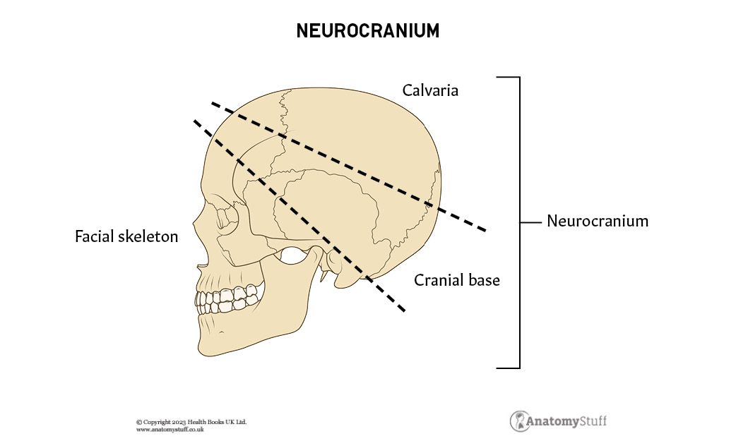
More specifically, the neurocranium can be subdivided into the cranial roof (calvaria) and skull base. The calvaria is composed of the frontal, parietal, temporal and occipital bones, and the skull base is composed of the sphenoid and ethmoid bones, and parts of the frontal, temporal, and occipital bones.
Frontal bone
The frontal bone is a thick, unpaired structure that forms the anterior and superior portion of the skull. As the name suggests, it protects the frontal lobe of the brain, as well as forming the superior aspect of the orbit (a socket within the skull in which the eye and its associated structures are located – more information below).
The frontal lobe contains a small depression called the glabella which is the smooth part of the forehead, in between the eyebrows.
Temporal bone
The temporal bone is a paired structure located at the sides and bases of the skull. It can be divided into several different parts and processes:
• Squamous part
• Mastoid part
• Temporal part
• Petrous part
• Styloid process
• Zygomatic process
As well as protecting the brain, the temporal bone surrounds the middle and inner portions of the ear, and connects with the lower mandible (jawbone) to allow the mouth to open and close. Additionally, many cranial nerves pass over the temporal bone.
Parietal bone
The parietal bone is a paired structure that forms the majority of the sides and roof of the skull. It is large and thin, with four borders and angles.
Occipital bone
The occipital bone is a trapezoidal-shaped bone forming the base of the skull. It is the most posterior cranial bone and it borders both parietal bones. Where they meet is known as the lambdoid suture (discussed in more detail below).
Sphenoid bone
The sphenoid bone resembles the shape of a butterfly and it is located in the middle of the skull between the frontal and temporal bones. The superior surface of the sphenoid body contains some important bony landmarks, such as the sella turcica, which is a saddle-shaped depression where the pituitary gland is located.
Ethmoid bone
The ethmoid bone is a small, unpaired bone located between the bony orbits. It separates the nasal cavity from the brain and forms the lateral borders of the bony orbit.
Bony landmarks
The pterion is a bony landmark located at the junction of the frontal, sphenoid, parietal and temporal bone. It is the thinnest and weakest part of the skull, making it prone to injuries like a fracture. The anterior branch of the middle meningeal artery runs beneath it.
The asterion is an anatomical landmark located at the junction of the occipital bone, the temporal bone, and the parietal bone. It is a reliable landmark for surgical approaches.
Cranial Fossae
The skull base, which forms the floor of cranial cavity, can be subdivided into three distinct cranial fossae:
- Anterior cranial fossa
- Middle cranial fossa
- Posterior cranial fossa
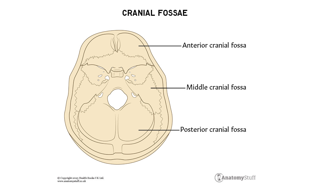
The anterior cranial fossa consists of the frontal, ethmoid, and sphenoid bones, the middle cranial fossa consists of the sphenoid bone and two temporal bones, and the posterior cranial fossa consists of the occipital bone and two temporal bones.
Foramina
In the skull base, there are numerous openings which allow important structures, such as cranial nerves and blood vessels, to pass from one region into another. These are referred to as cranial foramina.
Below is a brief list of the most important foramina and associated structures.
Foramina in the anterior cranial fossa:
| Foramina | Associated structures |
| Cribriform foramina | · Olfactory nerve (CN I) |
Foramina in the middle posterior fossa:
| Foramina | Associated structures |
| Optic canal | · Optic nerve (CN II)
· Ophthalmic artery |
| Superior orbital fissure | · Oculomotor nerve (CN III)
· Trochlear nerve (CN IV) · Abducens nerve (CN VI) · Ophthalmic division of trigeminal nerve (CN V1) |
| Foramen rotundum | · Maxillary branch of trigeminal nerve (CN V) |
| Foramen ovale | · Mandibular branch of trigeminal nerve (CN V) |
| Foramen spinosum | · Middle meningeal artery |
| Internal acoustic meatus | · Facial nerve (CN VII)
· Vestibulocochlear nerve (CN VIII)
|
Foramina in the posterior cranial fossa:
| Foramina | Associated structures |
| Hypoglossal canal | · Hypoglossal nerve (CN XII) |
| Foramen magnum | · Brainstem
· Spinal root of the accessory nerve (CN XI) · Vertebral arteries |
Sutures of the Skull
The bones of the skull are separated by very narrow junctions called sutures. They are a unique type of fibrous joint with a ‘fracture-like’ appearance. In children, sutures enable the skull to expand alongside the brain as it grows, and it closes around the age of 24.
The main sutures in the adult skull are:
- Coronal suture – fuses the frontal bone with the two parietal bones.
- Sagittal suture – fuses both parietal bones to each other.
- Lambdoid suture – fuses the occipital bone to the two parietal bones.
Fontanelles
In neonates, the incompletely fused suture joints give rise to membranous gaps between the bones, known as fontanelles (also called ‘soft spots’). Six fontanelles are present during infancy, with the two major ones being anterior and posterior fontanelles. As newborns grow, they close by a process called intramembranous ossification.
Summary of the anterior and posterior fontanelles:
| Fontanelle | Shape | Average closing time (after birth) | Conditions associated with delay in closure |
| Anterior | Diamond | 13-24 months | Down syndrome |
| Posterior | Triangular | 6-8 weeks | Hydrocephalus or congenital hypothyroidism |
Viscerocranium
The viscerocranium refers to the 14 bones that support the facial skeleton. There are six paired bones and two unpaired bones. They are also all unmoveable, besides the mandible.
The facial bones include:
• Zygomatic (paired) – diamond-shape bones that form the cheeks and the lateral walls of the orbits.
• Lacrimal (paired) – very small bones that form the medial walls of the orbits. They provide the groove for the tear ducts.
• Nasal (paired) – small, oblong bones located at midline of the face to form the bridge of the nose.
• Inferior nasal conchae (paired) – attached to the lateral wall of the nasal cavity and separates the middle nasal meatus from the inferior nasal meatus.
• Palatine (paired) – L-shaped bones that form the hard palate with the maxillary bones.
• Maxilla (paired) – central bones that form the dominant portion of the face.
• Vomer – thin, unpaired bone that divides the nasal cavity.
• Mandible (also known as the jaw) – the strongest and largest bone of the face. It forms the lower jaw and contains the lower teeth. It moves up and down during actions like speaking and chewing.
The maxilla is often considered the most important bone in the face, because it forms such a large proportion of it. It connects with surrounding facial structures through four processes:
• Alveolar
• Frontal
• Zygomatic
• Palatine
The maxilla bone separates the nasal and oral cavities, forms the upper jaw, and contains the maxillary sinuses.
The orbit
The orbits are two bony structures also known as the eye sockets. These paired, cone-shaped cavities contain the eyeball and its associated structures, such as muscles, nerves and blood vessels. Any space within the orbit that is not occupied is filled with orbital fat.
Seven bones contribute to the walls of the orbit, including:
• Maxilla
• Zygomatic
• Lacrimal
• Palatine
• Frontal
• Ethmoid
• Sphenoid
There are three several openings within the orbit, which allow important structures to pass. These include the optic canal, superior orbital fissure and inferior orbital fissure (discussed above).
Paranasal Sinuses
The paranasal sinuses are paired, hollow spaces located within several bones of the skull, more specifically the frontal bone, ethmoid bone, and sphenoid bone. These air-filled cavities open into the nose and their functions include supporting the immune system and humidifying inhaled air.
Embryology
Skull development begins around 23 to 26 days of gestation and derives from the neural crest and mesoderm. The facial bones, along with the frontal, ethmoid, and sphenoid bone, derive from the neural crest. The parietal and occipital bones originate from the mesoderm. The temporal bones derive from both the mesoderm and neural crest.
Refresher:
A germ layer is a group of cells in the embryo that differentiate into different tissues and organs. The three germ layers are the endoderm, the ectoderm and the mesoderm.

Related Products
View All


AnatomyStuff
Skull Anatomy Revision Worksheets (Interactive & Printable PDFs)
Now
£5.00
Inc VAT


