


![]()
Introduction
The spine, or vertebral column, is a skeletal structure composed of a collection of 33 vertically stacked bones called vertebrae. Each vertebra is separated by intervertebral discs, which form the fibrocartilaginous joint that anchor the vertebrae together and enable movement of the vertebral column.
Function of the Spine
The spine is the central support structure of the body and supports the weight of the upper torso, enabling the distribution of the weight to the lower limbs. It allows the attachment of various muscles and ligaments, therefore facilitating flexible motion and stability and is important in maintaining good posture. Aside from its structural support, it houses and protects the spinal cord within the vertebral cavity.
Embryology and Development of the Spine
The precursor population of cells responsible for spinal development are the somites. These are axial structures found in the paraxial mesoderm during early development. It is the medioventral region of the somites, known as the sclerotome, which eventually develops into the vertebral column.
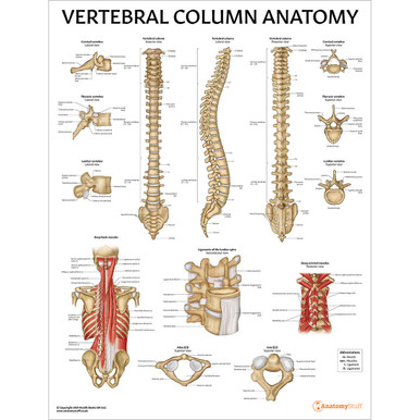
Structure of a Typical Vertebrae
The general structure of a vertebra consists of a vertebral body, a vertebral arch at the lateroposterior aspect, and a vertebral foramen. The vertebral body is the vertebra’s weight-bearing region and can be identified at the anterior aspect of the spinal column. It is continuous with the vertebral arch to form an enclosure, the vertebral foramen, and this opening provides a passage for the spinal cord to run through.
The vertebral arch is made up of processes that provide distinct attachment sites for muscles and ligaments. Extending laterally towards the back on each side of the vertebra are transverse processes. At the thoracic region, the transverse processes are the site of articulation for the ribs. Medial to the transverse processes is the spinous process, which extends posteriorly and provides an attachment for muscles and ligaments. The transverse processes are connected to the vertebral body via pedicles and to the spinous process via laminae. The vertebral arch consists of 2 pedicles and 2 laminae. At the superior aspect of the pedicles, further articular processes enable joints to be formed to the vertebra above.
Structure of the Vertebral Column
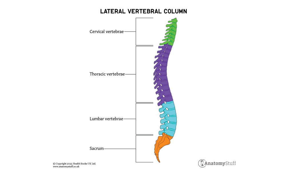
The vertebral column is part of the axial skeleton and is divided into 5 regions: the cervical, thoracic, lumbar, sacral, and coccygeal vertebrae. There are 7 cervical vertebrae, 12 thoracic vertebrae, 5 lumbar vertebrae, 5 sacral vertebrae and a range of 3 to 5 coccygeal vertebrae. In adulthood, the sacral and coccygeal vertebrae fuse to form a single bone, known as the sacrum and coccyx, respectively.
Movement of the spine is permitted by the presence of joints throughout the vertebral column, e.g., the joints of the vertebral bodies and joints of the vertebral arches. Looking at the spinal column from the lateral aspect, 2 curvatures, primary and secondary, can be identified. The primary curvatures (also referred to as the kyphotic curves) are convex and curve outwardly. This is seen at the thoracic and sacrococcygeal vertebrae. The secondary curvatures (known as lordotic curves) are concave and curve inwardly. This is seen at the cervical and lumbar vertebrae.
Throughout the spinal column, each adjacent vertebra is separated by intervertebral discs (IVD), fibrocartilaginous cushions that anchor 2 adjacent vertebrae to one another. The IVDs are shock-absorbing cushions which absorb stress that could incur during movement. It comprises a central nucleus pulposus, surrounded by a series of ringed fibrocartilage layers consisting of type 1 and 2 collagen, known as the annulus fibrosus. The nucleus pulposus is a gelatinous centre comprised of a mixture of water, collagen, and proteoglycans. IVDs are absent between the C1 and C2 vertebrae, as well as between the sacrum and coccyx.
Cervical Spine
The cervical spine is the uppermost part of the spinal column and is located within the neck region. It consists of 7 vertebrae, labelled C1 through to C7. The vertebrae at C3 to C6 follow the structure of a typical vertebra; however, C1, C2 and C7 are atypical, with distinguishable features.
The first cervical vertebra (C1) is also known as the atlas and is unique because it lacks both a vertebral body and spinous process. At the superior surface, 2 depressions, the superior articulating facets, can be identified. These facets articulate with the occipital condyles of the occipital bone at the rear of the cranium and form the synovial condyloid joint. The inferior articulating facets are 2 depressions found on the inferior surface that articulate with the superior articulating facets of C2 to form the synovial pivot joint. Together, these joints enable pivotal and rotational movements of the head.
The axis (C2) is distinguished by the presence of the odontoid process. This is a tube-like protuberance projecting cranially from the vertebral body and acts as a pivot point for C1. The C7 vertebra follows a similar structure to a typical vertebra but differs because it has a long, single spinous process.
From C3 to C6, the shape follows that of a typical vertebra and increases in size caudally to enable the increasing support of the weight of the head. On the transverse processes, openings known as the foramen transversarium (or transverse foramina) allow the passage of vertebral arteries. This is unique to the cervical vertebrae. The main function of the cervical spine is to support the head, protect surrounding spinal cords and allow flexible movement of the head.
Thoracic Spine
The thoracic spine is located between the cervical and lumbar vertebral segments. It comprises 12 vertebrae stacked on top of one another, labelled T1 to T12, each of which has a corresponding pair of ribs. The vertebrae from T2 to T8 are described as typical, whereas T9 to T12 and T1 are atypical. Regarding size, each thoracic vertebra is larger than the cervical vertebrae but smaller than the vertebrae found in the lumbar region. This enables greater support of the body’s weight, especially when standing, due to gravitational effects.
A typical thoracic vertebra is heart-shaped, with a long spinous process and circular vertebral foramen. The laminae are usually broad and overlap with the lower vertebra. A distinguishing characteristic is the presence of superior, inferior, and transverse costal facets on either side of the body. The superior costal facets articulate with the head of the numerically corresponding rib, and the inferior costal facets articulate with the head of the rib below. The transverse costal facet articulates with the rib’s tubercle at that level. Superior and inferior articular processes can also be identified, which articulate with the upper and lower vertebra.
The first thoracic vertebrae (T1) is atypical because, although it has a complete superior costal facet, the inferior costal facet is incomplete and is known as the demi facet. Only 1 costal facet can be identified on each side of the vertebral bodies of T9 to T12, which is complete in all vertebrae, apart from T9, which is a demi facet.
Due to its articulation with the ribs, the thoracic spine aids in the protection of important organs, such as the heart and lungs. It provides flexibility to the thoracic cavity, which is important in respiration, especially during inspiration and expiration. Additionally, it protects the spinal cord and supports the chest and abdomen.
![]()
Lumbar Spine
The lumbar spine is found in the lower region of the spinal column, between the thoracic and sacral vertebrae, and consist of 5 moveable vertebrae, labelled L1 to L5, or L6 in some individuals. Anatomically, it is positioned inferior to the ribcage and superior to the pelvis.
This region is largely responsible for bearing the upper body’s weight and is maintained by the intervertebral discs, muscles, and ligaments. Ligaments, such as the interspinous and supraspinous ligaments, are present throughout the vertebral column, but unique to the lumbar spine is the iliolumbar ligament, which functions to strengthen the lumbosacral joint (between the L5 and S1 vertebrae).
The lumbar vertebrae are large, kidney-shaped, thick structures of dense bone that are bigger than vertebrae from other parts of the vertebral column. L1 to L4 follows the structure of a typical lumbar vertebra, and L5 is atypical. The transverse diameter of the vertebral body is wider than the anterior-posterior diameter. The vertebral foramen is triangular, and the foramen transversarium is absent on the transverse processes and facets on the lateral surfaces of the body.
Generally, the lumbar vertebrae consist of long transverse processes, articular processes, short spinous processes, accessory processes, and mamillary processes. The accessory processes are the small elevations at the posteroinferior aspect of the root of each transverse process. The mamillary process is an elevation located at the posterior border of the superior articular process. Both the accessory and mamillary processes are sites of attachment for deep back muscles. For example, the mamillary process provides attachment for the multifidus and medial intertransversarii muscles.
The well-defined superior and inferior articular processes project upwards and downwards from the junctions of the pedicles and laminae. The superior processes are concave and face medially, but the inferior processes are concave and face outwards towards the superior processes of the vertebra below. This creates resistance when the lower spine is twisted.
At the atypical lumbar vertebra (L5), the vertebral body is the largest of them all, and the transverse and spinous processes are shorter and thicker. The anterior surface of the body is more extensive than the posterior surface and is responsible for the sharp lumbosacral angle. A key function of the lumbar spine is to support and stabilise the upper body, as it is the centre of the body’s balance. The connection and interaction of the lumbar spine to certain muscles and ligaments enable truncal movements, such as flexion, extension, rotation, and side bending.
Sacrum
The sacrum is found at the inferior region of the spinal column and is composed of 5 sacral vertebrae (S1 to S5) with an inverted, triangular, and concave shape. Although it is initially 5 separate vertebrae, this fuses into one sacral bone during adulthood. As a result of this fusion, there are transverse ridges or lines that can be seen between each vertebra.
At the superior aspect, the superior articular processes of the sacral vertebra (S1) articulate with the 5th lumbar (L5) vertebra and creates the synovial plane joints. This forms the lumbosacral curve that is subject to great amounts of stress and is prone to injuries. The lateral surface of the sacral body articulates with the auricular surface of the ilium (the largest part of the hip bone) via a sacroiliac joint. This forms the pelvic girdle and provides stability and strength to the pelvis architecture. The shape of the sacrum varies between genders. In females, it is shorter and broader, thus increasing the overall size of the pelvic cavity and aiding in pregnancy.
Medial to the superior articular processes is an opening known as the sacral canal. This is a continuation from the vertebral canal and contains the sacral nerves. The sacral nerves enter through the sacral canal and exit via the sacral foramina. The foramina are openings found lateral to the transverse ridges on the ventral surface and lateral to the median crests on the dorsal surface.
On the posterior surface, there are 3 crests, the median, intermediate and lateral sacral crests. The median sacral crests are the remnants formed from the fusion of the spinous processes and are attachment sites for the supraspinous ligament. The intermediate sacral crest allows attachment of the posterior sacroiliac ligaments. The fusion of the transverse processes form the lateral sacral crest and provide the site of attachment for the sacrotuberous and sacroiliac ligaments.
At the caudal end of the sacrum, a gap known as the sacral hiatus is formed by the 2 sacral cornua. This articulates with the coccygeal cornua of the coccyx bone below. The inferior end of the sacrum is the apex. The apex articulates with the coccyx bone and forms a joint known as the sacrococcygeal symphysis.
Coccyx
The inferior portion of the vertebral column is made up of the coccyx and is a triangular bone made up of approximately 3 – 5 fused vertebrae (Co1 – Co5). In some adults, not all vertebrae are fused together. The coccyx bone is an irregular bone of the axial skeleton and is commonly referred to as the ‘tailbone’. The shape of the coccyx varies between genders, where the male coccyx is curved at the front, and the female coccyx is straighter and has greater flexibility.
On the superior region of the posterior surface, there are 2 tubercle projections known as the coccygeal cornua, which articulate with cornua of the sacrum above. The base of the coccyx forms an amphiarthrodial joint, called the sacrococcygeal symphysis, with the sacral apex. This joint enables only slight movements and is limited to passive flexion and extension, which can arise due to intra-abdominal pressure.
On either side of Co1 are transverse processes which extend outwards laterally and provide a passage for the fifth sacral nerve. The apex is found at the terminal end of the coccyx and acts as an attachment site for various muscles, tendons, and ligaments, e.g., the tendon of the external anal sphincter. The lateral borders of the coccyx are also attachment sites for muscles, such as the gluteus maximus and coccygeus, in addition to important ligaments, including the sacrotuberous and sacrospinous ligaments.
Overall, the coccyx functions to provide stability and weight-bearing support to an individual, especially at a seated position. It further supports the surrounding pelvic organs.

Related Products
View All

save
40%
AnatomyStuff
Budget Cervical Spine Model
Now
£13.20
Inc VAT
Was
£22.00
Inc VAT

save
30%
AnatomyStuff
Spine Anatomy Poster / Worksheet (Interactive & Printable PDF)
Now
£3.50
Inc VAT
Was
£5.00
Inc VAT


