


![]()
Introduction
The thorax refers to the part of our body between the neck and the abdomen. The space surrounded by the thoracic wall is called the thoracic cavity. Multiple organs are contained in the thoracic cavity. The thorax plays a central role in the ventilation process. It always provides adequate protection for organs of the respiratory and cardiovascular systems.
Thoracic Wall
The thoracic wall plays a vital role in inspiration. It resists overinflation of the lungs when air is breathed in. It is also the site of attachment of the upper limbs and many muscles of varying origins, for example, muscles of the neck, upper limb, back, abdomen and respiratory muscles.
Boundaries of the thoracic wall include:
• Anterior – Sternum (this bone is divided into three parts (from superior to inferior): manubrium, body, and xiphoid process)
• Posterior – Twelve thoracic vertebrae
• Lateral – Twelve ribs on either side of the thoracic cavity
• Superior – Superior thoracic aperture
An opening in the thoracic cage that allows vessels from the neck to travel into the thorax. These vessels include the trachea, oesophagus, nerves etc. Bound by the margins of vertebrae T1, the medial margin of the first rib on either lateral side and the manubrium
• Inferior – Inferior thoracic aperture
An opening in the thoracic cage that allows vessels from the thorax to travel into the abdomen. Much larger than the superior thoracic aperture. The diaphragm marks the end of the inferior thoracic aperture as it separates the thoracic and abdominal cavities. Bound by the margins of vertebrae T12, margins of the twelfth rib on either lateral side, the cartilaginous ends of ribs 7-10 (anterolaterally) and the xiphoid process.
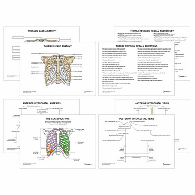
Bones of the Thoracic Wall
The thoracic cage consists of twelve pairs of ribs on either side. Each rib is attached to the sternum (could be directly or indirectly) through costal cartilages. All ribs attach posteriorly to the thoracic vertebrae. Ribs are categorised as curved, flat bones. They are further classified based on their attachments; and whether they are typical or atypical.
True, false and floating ribs
The first to seventh ribs are known as true ribs – this is because they attach directly to the sternum through their own costal cartilages.
The eighth to tenth ribs are known as false ribs – this is because they attach indirectly to the sternum through the costal cartilages of the ribs above them.
The eleventh to twelfth ribs are known as floating ribs – this is because, unlike the other ribs, they have no anterior connection to the sternum.
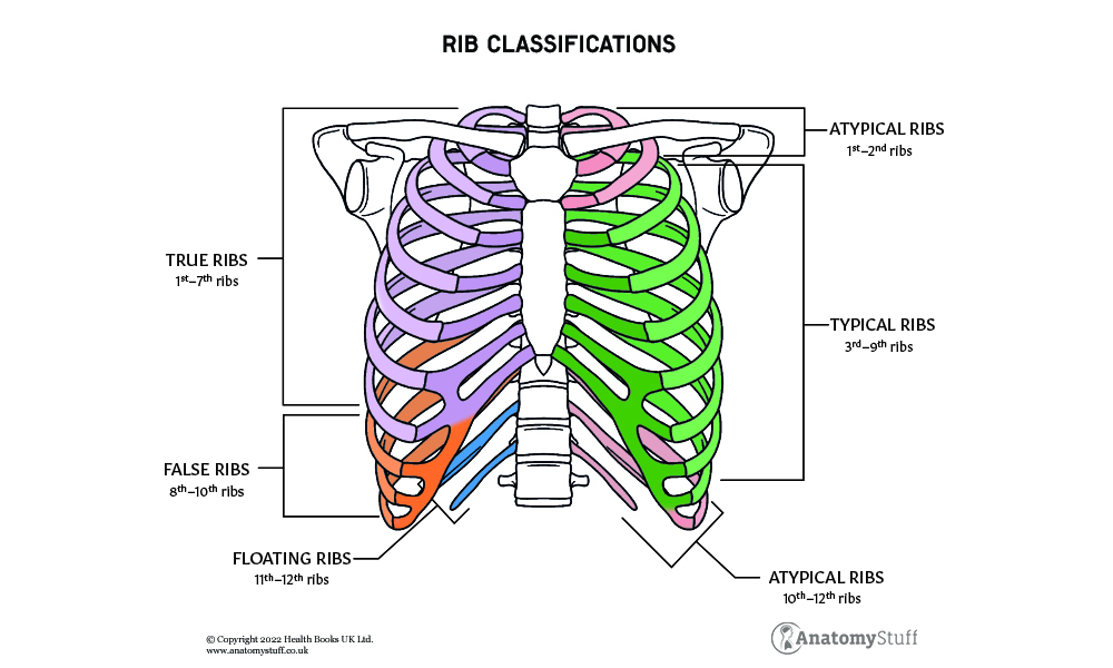
Typical and atypical ribs
Typical ribs have features that are common to all the ribs in this category. These features include:
Head: this is present at the posterior end of the ribs. It has two facets that are separated by the crest of the head. The superior facet of the rib is the site of articulation for the inferior demi facet of the anatomically superior vertebrae. The inferior facet of the rib articulates with the superior demi facet of the numerically corresponding vertebrae. E.g., the Superior facet of rib 3 attaches to the inferior demi facet of T3, and the inferior facet of rib 3 attaches to the superior facet of T4.
Neck: this is a flat surface of bone between the head and the tubercle.
Tubercle: this is a bony projection that contains an articular surface and a non-articular surface. The smooth articular surface articulates with numerically corresponding transverse process of thoracic vertebrae. The rough non-articular surface is the site of attachment for the costotransverse ligament, which also attaches to the transverse process of the vertebrae.
Shaft: this is the flat, curved part of the rib. At a point known as the costal angle on the shaft, the rib begins to turn anterolaterally. Along the inferior margin of the shaft runs the costal groove where veins, arteries and nerves are contained.
Atypical ribs have distinct features, unlike typical ribs.
First rib: this is the shortest and widest of all ribs
Unlike the typical ribs, the head of the first rib only has one true facet that attaches to the T1 vertebrae. It has a tubercle for articulation with the transverse process. There are two grooves in the shaft of the rib that are separated by the scalene tubercle. The subclavian vein causes the anterior groove, and the posterior groove is caused by the subclavian artery. The scalene tubercle acts as a site of attachment for the anterior scalene muscle of the neck.
Second rib: nearly twice the length of the first rib
Most features of the second rib conform to those of typical ribs. However, it has formations along its body for attachment of the serratus anterior muscle and posterior scalene muscle.
Tenth, eleventh and twelfth ribs: similar to the first rib
All these ribs have one true facet on their head for attachment to the numerically corresponding vertebrae. Ribs eleven and twelve have no necks or tubercles and are anteriorly positioned.
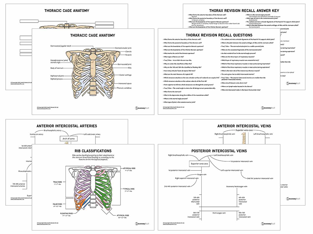
The sternum is a wide bone that contains the sites of attachment for all the true ribs (1-7). It has three distinct processes:
Manubrium (Superior)
Contains the jugular (suprasternal) notch along the midline. On either side, it contains fossa for the sternoclavicular joint and an attachment site for the first rib.
Body of sternum (Middle)
Along the lateral margins are sites of articulation of the costal cartilages of ribs two to seven. The manubrium is attached to the body of the sternum through the manubriosternal joint, also known as the sternal angle, which is clinically very relevant.
Xiphoid process (Inferior)
This is the smallest part of the sternum. It is formed as cartilage and ossifies over time in adults.
![]()
Joints and Ligaments of the Thoracic Wall
The thoracic cage consists of multiple bones that are bound by joints. The stability of these joints is vital for breathing; however, the movements of these joints are relatively minimal.
The joint between the rib and thoracic vertebrae (allows for the increasing volume of the thorax during breathing)
Costovertebral joint (synovial plane joint) – the head of the rib articulates with a demi facet present on the thoracic vertebrae
• Associated ligaments: radiate and intra-articular ligaments of the head of the rib.
Costotransverse joint (synovial joint) – tubercle present along the body of the rib articulates with the transverse proves of the thoracic vertebrae
• Associated ligaments: lateral and superior costotransverse ligaments.
Sternocostal joint – the joint between the costal cartilages of the first to seventh ribs and the sternum
• 1st sternocostal joint (primary cartilaginous joint) – articulation with the manubrium.
• 2nd to 7th sternocostal joint (synovial plane joints) – articulation with the body of the sternum.
• Associated ligaments: anterior and posterior radiate sternocostal.
Sternoclavicular joint (saddle synovial joint) – the joint between manubrium and clavicle on either side
• Fibrocartilaginous discs are present to allow for shock absorption when an injury occurs.
• Associated ligaments: anterior and posterior sternoclavicular ligaments and costoclavicular ligaments.
Manubriosternal joint – the joint between the manubrium and body of the sternum
• Also known as the sternal angle, which is present at the T4/5 vertebral level.
Xiphisternal joint – the joint between the body of the sternum and the xiphoid process
• The xiphisternal joint and the manubriosternal joint fuse together as people age.
Intercostal Spaces
Spaces between ribs are known as intercostal spaces and have multiple structures present. These structures include muscles, veins, arteries, and nerves that aid in the respiratory process and provide stability to the thoracic wall.
Intercostal veins, arteries and nerves run through the costal groove along the inferior margin of a rib. In each space, from superior to inferior: vein, artery, and nerve. Usually, the nerve isn’t protected by the costal groove; therefore, it is at risk of damage during clinical procedures (especially a chest drain during pneumothorax). This bundle of vessels is present between the innermost intercostal muscle and internal intercostal muscle
Respiratory muscles allow expansion of the thoracic cage to allow for efficient respiration. There are three layers of respiratory muscles between the ribs:
External intercostal muscle
This muscle runs anteroinferior from the superior rib to the inferior rib. It allows for the ribs to move superiorly and is especially active during inspiration.
Internal intercostal muscle
This muscle runs in an opposite direction to the external intercostal muscle. It allows for the rib to move inferiorly and is most active during expiration.
Innermost intercostal muscle
This muscle runs in a similar direction and has a similar function to the internal intercostal muscle.
Transversus thoracis
This muscle runs from the costal cartilages of the second to sixth ribs to the body of the sternum/ xiphoid process. The main role of this muscle is to depress the costal cartilages.

Related Products
View All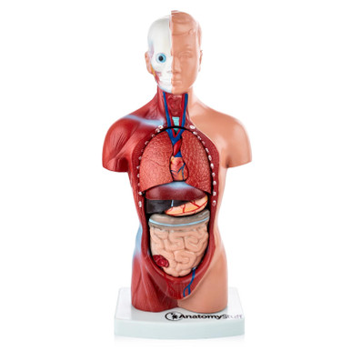

save
30%
AnatomyStuff
Lung Anatomy Poster / Worksheet (Interactive & Printable PDF)
Now
£3.50
Inc VAT
Was
£5.00
Inc VAT
AnatomyStuff
Thorax Anatomy Revision Worksheets (Interactive & Printable PDFs)
Now
£5.00
Inc VAT


