


Introduction
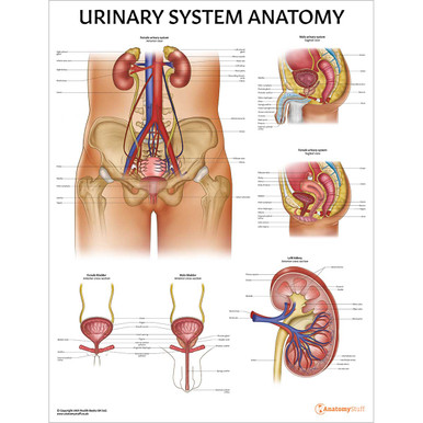
The urinary system consists of the kidneys, ureters, bladder, and urethra. These organs work together to produce, store and eliminate waste from the body in the form of urine. There are many common disorders involving these organs, such as tract infections (UTIs). UTIs can be divided into lower tract infections (involving the urethra and bladder) or upper tract infections (involving the ureters and kidneys) which are usually more serious. This page will discuss the anatomy of the urinary system as well as clinically relevant conditions such as UTIs and bladder cancer.
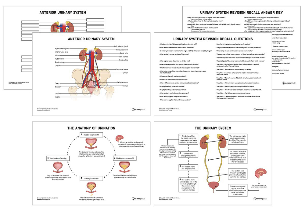
Summary of the Urinary System
As discussed above, the urinary system consists of the kidneys, ureters, bladder, and urethra. The kidneys remove a substance called urea from the blood through tiny filtering units called nephrons. Along with water and other waste substances from the blood, this forms urine. The ureters then transport the urine from the kidneys to the bladder. Both ureters pierce the wall of the bladder in an oblique manner (slanted position), preventing the reflux of urine back up to the kidneys.
The role of the bladder is to store urine before disposal by urination. The average capacity of a functional bladder is between approximately 300 to 400ml, although this changes with age. When the bladder is full, stretch receptors in the bladder wall trigger the micturition (urination) reflex, and the urine leaves the body through a hollow tube called the urethra.

Kidneys
Function
The kidneys, which are oval in shape, perform many life-sustaining functions. They are responsible for filtering excess water, salts, and waste products from the blood while returning nutrients and other vital substances back to the bloodstream.
Location
The kidneys are paired retroperitoneal organs (meaning they have peritoneum on their anterior side only). They are located on either side of the spine at the level of T12-L3 vertebrae, with the right kidney situated slightly lower due to the presence of the liver above.
Anatomy
On the medial border of the kidney, there is a vertical, concave slit called the renal hilum. This is the entry and exit point for important structures like the renal arteries and veins, lymphatic vessels, nerves and ureters.
The renal medulla, renal cortex and renal pelvis are the three main internal regions found in the kidneys.
The renal medulla is the inner part of the kidney containing the majority of the length of the nephrons. There are approximately 1 million nephrons per kidney, and they are responsible for producing urine.
The urine produced by the nephron flows to the calyces, which is where urine collection begins. The urine then flows into the renal pelvis, and from there, it flows into the ureter.
Essentially, the calyces and renal pelvis act as a reservoir for urine received from the nephron (filtering unit). The urine is then funnelled into the ureter.
Blood supply
The kidneys are supplied with blood via the renal arteries, which arise from the lateral aspect of the abdominal aorta at the level of L1-L2 vertebrae. They enter the renal hilum to supply the kidneys. Interestingly, there are many variations of renal artery anatomy, which is very important to consider during renal surgery or transplantation.
Most commonly, there are two renal arteries (right and left), with the right one being slightly longer than the left. However, people may also have additional arteries supplying the kidney, namely the hilar, upper polar and lower polar arteries.
Ureters
Function
The ureters propel urine from the kidneys to the bladder. The muscular layer of the ureters is responsible for the peristaltic activity that pushes the urine along, and the oblique positioning of the ureters as they enter the bladder (slanted) prevents the reflex of urine.
Location
The ureters begin at the ureteropelvic junction (UPJ) of the kidneys. They then travel inferiorly inside the abdominal cavity, passing over the psoas muscle and entering the bladder on the posterolateral wall. These paired narrow muscular ducts are roughly 30cm long.
Anatomy
Along each ureter, there are three narrow sections; the ureteropelvic junction (where the kidneys meet the ureter), the crossover of the common iliac arteries and the ureterovesical junction (where the ureters enter the bladder). These are common sites for ureteric stones to become trapped, causing severe loin pain and haematuria (blood in the urine).
The ureter can be divided into abdominal and pelvic segments. The abdominal ureter is the segment of the ureter that extends from the renal pelvis to the iliac vessels. The pelvic ureter extends from the iliac vessels to the bladder.
Surgical significance
Clinically, the ureters are highly significant as they are located close to many other major vessels and organs. For example, in females, the ovarian and uterine arteries lie very near to the ureters, and surgeons must therefore be careful to avoid accidental injury during an oophorectomy or hysterectomy.
Blood supply
Several different arteries supply the ureter. The upper part receives its blood supply from the renal artery and branches of the gonadal artery and aorta. The middle part of the ureter is supplied by the common iliac and gonadal arteries, and the distal part is supplied by the uterine and superior vesical arteries.
Interestingly, the ureter receives blood supply from both medial (abdominal ureter) and lateral (pelvic ureter) aspects. This is important to consider during urological surgery.
![]()
Bladder
Function
The bladder receives urine through the ureters. It stores this waste product until the central nervous system initiates urination. Parasympathetic stimulation encourages micturition, while sympathetic stimulation prevents it.
Location
The bladder is a triangle-shaped organ located in the lower abdomen. It sits in the lesser pelvis when empty and extends into the abdominal cavity when full.
Anatomy
The bladder is anatomically divided into four parts: the apex (top part), body (main portion between the apex and the fundus), fundus (base) and neck (narrow part connecting the bladder to the urethra).
Urine enters the bladder through the left and right ureters, and these orifices are marked by the trigone, which is a triangular area located within the fundus.
The bladder wall is made up of detrusor muscle composed of longitudinal and circular smooth muscle fibres. This muscle plays a key role in the storage and emptying of urine- it contracts during urination to push the urine into the urethra and relaxes to hold and store the urine.
Blood supply
In males, the superior and inferior vesical arteries supply the bladder. These are branches of the internal iliac artery. In females, the blood supply is slightly different, with the superior vesical and vaginal arteries supplying the bladder. Venous drainage corresponds to arterial supply, and together these veins form the vesical venous plexus.
Urethra
Function
The urethra transports urine from the bladder to the exterior of the body. The urethra is held closed by the external and internal urethral sphincters. When these muscles contract, urination stops.
Location
The male’s urethra originates at the bladder neck and terminates at the urethral meatus on the glans penis. In females, the urethra also originates at the bladder neck but opens into the vulva vestibule, inferior to the clitoris and superior to the vaginal opening.
Anatomy
In males, the urethra is approximately 20cm long. The urethra can be divided into three parts: prostatic, membranous and penile (also known as bulbous). It also transports semen for ejaculation.
In females, the urethra is approximately 5cm long. This is clinically relevant because it means that females are more likely to develop urinary tract infections (UTIs) due to the shorter length of the urethra. This is because bacteria have less distance to travel before reaching the bladder or kidneys to cause infection.
There are also two muscular sphincters located in the urethra, the internal and external urethral sphincters. The internal urethral sphincter is under autonomic control (involuntary), and the external urethral sphincter is under voluntary control.
Blood supply
The arterial supply to the male urethra is via several different arteries; the prostatic urethra is supplied by the inferior vesical artery, the membranous urethra is supplied by the bulbourethral artery and the penile urethra is supplied directly by branches of the internal pudendal artery.
Clinical relevance
Pathologies affecting the urinary system
Many disorders affect the organs of the urinary system, such as urinary tract infections, kidney stones, urinary incontinence and bladder cancer.
Urinary tract infections (UTIs) are very common (particularly amongst women) due to the shorter length of the urethra, as discussed above. UTIs involve infection in the bladder, causing cystitis (inflammation of the bladder), and the bacteria can also travel to the kidneys, causing pyelonephritis.
The primary source of bacteria causing UTIs is the faeces, as it travels easily from the anus to the urethral meatus.
Symptoms of lower UTIs include dysuria (pain during urination), discomfort and confusion in elderly patients. In upper UTIs, patients may also have a fever and back pain, which can be key signs that the infection may have travelled up to the kidneys.
Bladder cancer is another important condition involving the urinary tract. Transitional cell carcinomas are the most popular type, and the main risk factors are smoking and increased age. Visible painless haematuria (blood in the urine) is a red flag symptom and warrants an urgent referral (always refer to the up-to-date NICE guidelines).
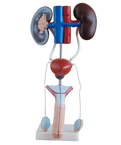
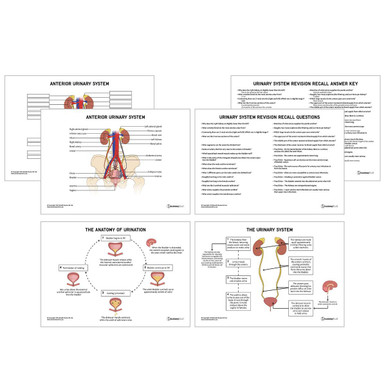
Take a look at our Urinary System Anatomy Chart for more visual learning.

Related Products
View All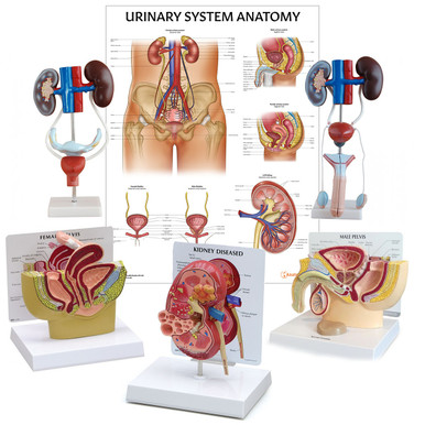
AnatomyStuff
Urinary System Anatomy Revision Worksheets (Interactive & Printable PDFs)
Now
£5.00
Inc VAT


