


![]()
Introduction
Pelvis
The pelvis is a basin-shaped structure connecting the trunk to the lower limb. It contains and supports many urogenital organs, including the distal region of the digestive system, the majority of the reproductive organs and part of the urinary system. There are also key adaptations between the male and female pelvis, reflecting the need for females to facilitate childbirth. Keep reading to learn all about the anatomy of the pelvis, including the different bones, muscles, ligaments, blood supply and innervation.
Bones
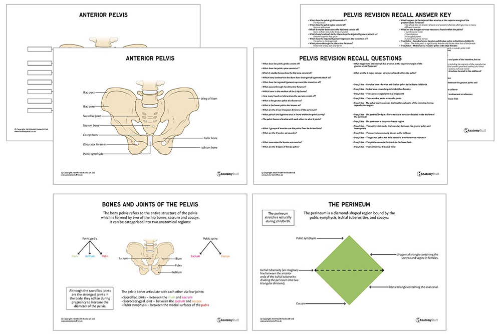
The bony pelvis refers to the entire structure of the pelvis, which is formed by two hip bones, sacrum and coccyx.
The pelvis can be categorised into two anatomical regions:
- Pelvic girdle –hip bone
- Pelvic spine – sacrum and coccyx
Each hip bone consists of three smaller bones: the ilium, ischium and pubis:
• The ilium is the largest and uppermost part of the hip bone. It is composed of a body and ala (wing). The body is the central portion located above the acetabulum and the wing is the larger, flatter portion which contains several important bony landmarks:
• Anterior superior iliac spine (often referred to as its abbreviated version of ASIS) = can be easily palpated and serves as a point of attachment for the inguinal ligament. This is a fibrous band which extends to the pubic tubercle. It is an important anatomical landmark because it notes the transition of the pelvis to the lower limb.
• Anterior inferior iliac spine = serves as a point of attachment for the rectus femoris and proximal part of the iliofemoral ligament.
• Posterior superior iliac spine
• Posterior inferior iliac spine
• The ischium is an L-shaped bone which forms the posteroinferior part of the hip bone. It consists of two parts – the body and the ramus. Its ramus joins with the inferior pubic ramus of the pubic bone, completing the obturator foramen, a through which the obturator artery, obturator vein and obturator nerve pass.
• The pubic bone is also known as the pubis. It is located at the anteroinferior aspect of the pelvis and is the smallest of the three hip bones. It consists of a body and two rami (superior and inferior), which meet medially at the body of the pubis. On the anterior aspect of the body is a bony thickening called the pubic crest, and on the lateral aspect of this crest is the pubic tubercle.
The sacrum is a thick triangular-shaped bone located below the last lumbar vertebra (L5). It consists of five fused vertebrae. The coccyx, commonly known as the tailbone, is below the sacrum.
Greater and lesser pelvis
The space within the pelvic bones is called the pelvic cavity, which can be divided into two parts:
- The greater pelvis, also known as the false pelvis, is located superiorly. It is considered to be part of the abdominal cavity and has little obstetric involvement or relevance. It supports the abdominal organs.
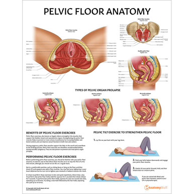
- The lesser pelvis, also known as the true pelvis, is located below. The pelvic floor is the inferior muscular layer of the true pelvic cavity which separates it from the perineal region (including the perineum) below.
The pelvic inlet marks the boundary between the greater pelvis and lesser pelvis. Its size is defined by its edge, the pelvic brim.
Perineum
The perineum is a diamond-shaped region bound by the pubic symphysis, ischial tuberosities, and coccyx. An imaginary line between the anterior ends of the ischial tuberosity divides it into two triangular divisions:
The urogenital triangle (which contains the urethra and vagina in females) and the rectal triangle (which contains the anal canal). The perineal body, a fibro-muscular structure located in the midline of the perineum, is located between these hiatuses. The perineum stretches naturally during childbirth.
Organs
The pelvic cavity contains urogenital organs, including the majority of the reproductive organs, some of the urinary system (urinary bladder, distal ureters, proximal urethra) and the distal part of the digestive tract (terminal sigmoid colon, rectum, and anal canal).
Joints
The pelvic bones articulate with each other via four joints:
• Sacroiliac joints – two synovial joints between the ala of the sacrum and the auricular surface of the ilium. It is the strongest joint in the body, although they soften during pregnancy to increase the diameter of the pelvis.
• Sacrococcygeal joint – a hinge joint between the sacrum and the coccyx. It allows flexion and extension of the coccyx during childbirth and defecation.
• Pubic symphysis – a cartilaginous joint between the medial surfaces of the pubic bones. Usually, there are no movements within this joint, except in pregnancy when the ligaments and cartilage soften.
![]()
Muscles
The pelvic floor refers to a group of muscles that support the organs in the pelvis. They can be divided into two main groups: levator ani muscles and coccygeus muscles. The levati ani muscle group includes:
• Pubococcygeus
• Puborectalis
• Iliococcygeus
They are innervated by branches of the pudendal nerve and the anterior ramus of S4.
The coccygeus, also known as ischiococcygeus, is a triangular-shaped sheet of muscle located posterior to the levator ani muscles in the pelvic floor.
Sex differences
There are apparent differences between the male and female pelvis. The adult female pelvis is usually broader, with a rounder pelvic inlet. The angle of the pubic arch is greater than 90 degrees, and these adaptations reflect the necessity to provide an adequate-size birth canal during childbirth. The male pelvis has a higher iliac crest with a longer, more narrow sacrum. In general, it is significantly heavier and thicker than that of the female.
Types
The shape of the female bony pelvis can be classified based on the shape of the pelvic inlet into four broad categories: gynecoid, anthropoid, android, and platypelloid.
The gynecoid pelvis (rounded shape) is known as the typical female type because it is adapted to facilitate pregnancy and childbirth. The android pelvis (heart-shaped) is often designated a male variant.
Age changes
The pelvis has been reported to change in shape with age. The most rapid period of growth is in the first year of life, and a stable rate of growth is attained after age 3 years until puberty.
Blood supply
The internal iliac arteries, also known as the hypogastric arteries, are branches of the common iliac arteries. They are approximately 4cm in size and provide blood supply to the pelvic organs, muscles and perineum.
The internal iliac arteries pass medially over the pelvic brim and continue downwards to reach the greater sciatic foramen. At the superior margin of the greater sciatic foramen, they divide into an anterior division and posterior division, which give rise to many different branches:
| Branches of the anterior division | Blood supply |
| Obturator artery | Pelvic muscles, ilium, head of femur, muscles of medial thigh, skin |
| Umbilical artery | Superior aspect of the bladder |
| Inferior vesical artery | Inferior aspect of the bladder, prostate gland* and seminal vesicles* |
| Vaginal artery | Vagina** |
| Uterine artery | Uterus** |
| Middle rectal artery | Distal part of the rectum |
| Internal pudendal artery | Perineum |
| Inferior gluteal artery | Gluteal muscles and hip joint |
| Branches of the posterior division | Blood supply |
| Iliolumbar artery – lumbar branch | Psoas major, quadratus lumborum and the posterior abdominal wall |
| Iliolumbar artery – iliac branch | Muscles and bone around the iliac fossa. |
| Lateral sacral arteries (superior and inferior) | Structures in the sacral canal and skin over the sacrum
|
| Superior gluteal artery | Muscles and skin of the gluteal region |
* Males only, ** Females only
Venous drainage is achieved primarily by the internal iliac vein. The internal iliac vein combines superiorly with the external iliac vein to form the common iliac vein, which then drains into the inferior vena cava.
Innervation
There are four major nervous structures found in the pelvis which supply the muscles of the pelvic floor and perineum, the gluteal region, pelvic viscera (including the urinary bladder, the distal end of the ureters, rectum and reproductive organs) and the lower limb. These structures are:
• Lumbosacral trunk
• Sacral plexus
• Coccygeal plexus
• Autonomic pelvic nerves
Lymphatic drainage
The lymphatic drainage of the pelvis is similar to the abdomen. Pelvic organs, like the female reproductive organs (uterus, cervix, and vagina), drain into the internal and external iliac nodes. These are found adjacent to the external, internal and common iliac arteries.
You can learn more with our lymphatic revision guide!
![]()

Related Products
View All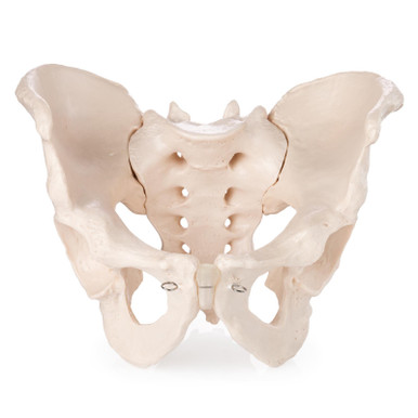

AnatomyStuff
Pelvis Anatomy Revision Worksheets (Interactive & Printable PDFs)
Now
£5.00
Inc VAT


