Written by: Liz Paton, MSc
Pharynx & Larynx Anatomy Overview
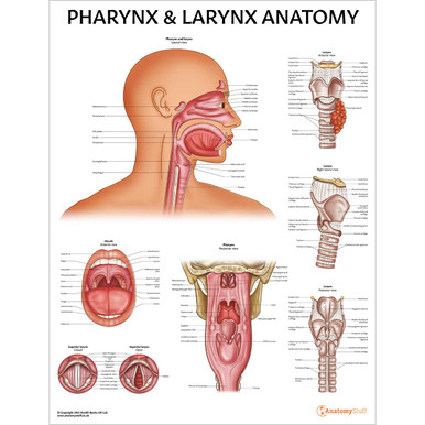
Anatomy of the Larynx
The pharynx, Greek for the throat, is situated behind the oral and nasal cavity and acts as the passageway from the oral and nasal cavities to the oesophagus and larynx. It is responsible for respiratory and digestive functions.
Anatomy of the Pharynx
The pharynx is made up of muscle, mucous membrane, submucosal connective tissue, lymphoid tissue and glands. It can be broken down into three main divisions: the nasopharynx, oropharynx and laryngopharynx.
The nasopharynx is the upper-most part of the pharynx found between the nasal cavity and soft palate. It forms the upper part of the respiratory system by passing inspired air from the nose to the larynx.
The oropharynx is located in the back of the mouth, between the oropharynx and laryngopharynx. It aids with digestion by allowing us to breathe while we masticate (chew) our food.
The laryngopharynx, also known as the hypopharynx, is the lowest portion of the pharynx, located just beneath the oropharynx. This is the point where the pharynx meets the larynx and where food, water and air pass through.
The Larynx
The larynx (the Greek word larynges meaning “the upper windpipe”) is located in the anterior part of the neck. The larynx is also often referred to as the windpipe and voice box. It is responsible for air passage, producing sound and swallowing food.
Anatomy of the Larynx
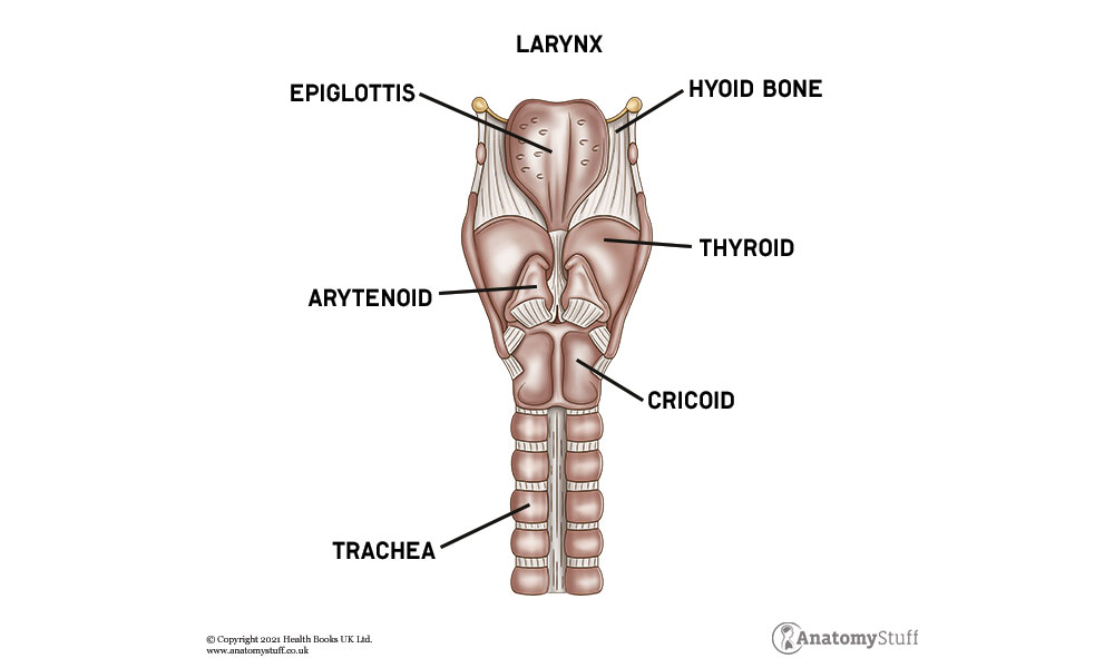
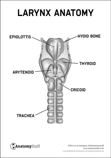
The larynx is a hollow structure, made up of muscle, ligaments and cartilage. It has three unpaired cartilages, the thyroid, cricoid and epiglottis, and three paired cartilages, the arytenoid, cuneiform and corniculate.
The thyroid cartilage is the largest structure of the larynx that forms what is known as the “Adam’s apple”. It is made up of hyaline cartilage and serves as an attachment for several small muscles. The cricoid cartilage sits inferior to the thyroid. It is the only continuous ring around the trachea. The epiglottis lays just above the larynx and acts as a lid, preventing food and liquid from entering the pharynx.
The arytenoid cartilages are a pair of pyramid-shaped structures to which the vocal cords attach, and are essential for producing sound. The cuneiform cartilages are a pair of wedge-shaped cartilages that support the vocal folds. The corniculate cartilages are a pair of horn-like structures, just below the cuneiform cartilages, that articulate with the arytenoid cartilages.